Human/Mouse/Rat Syntaxin 7 Antibody
R&D Systems, part of Bio-Techne | Catalog # AF5478

Key Product Details
Species Reactivity
Validated:
Cited:
Applications
Validated:
Cited:
Label
Antibody Source
Product Specifications
Immunogen
Asn21-Glu187
Accession # O15400
Specificity
Clonality
Host
Isotype
Scientific Data Images for Human/Mouse/Rat Syntaxin 7 Antibody
Detection of Human Syntaxin 7 by Western Blot.
Western blot shows recombinant human Syntaxin 12, 16, 1A, 1B2, 5, 6, 7, 8, and 1B1 (5 ng/lane). PVDF membrane was probed with 1 µg/mL Sheep Anti-Human/Mouse/Rat Syntaxin 7 Antigen Affinity-purified Polyclonal Antibody (Catalog # AF5478) followed by HRP-conjugated Anti-Sheep IgG Secondary Antibody (HAF016). A specific band for Syntaxin 7 was detected at approximately 29 kDa (as indicated). This experiment was conducted under reducing conditions and using Immunoblot Buffer Group 1.Detection of Human, Mouse, and Rat Syntaxin 7 by Western Blot.
Western blot shows lysates of BJAB human Burkitt's lymphoma cell line, MCF-7 human breast cancer cell line, C2C12 mouse myoblast cell line, BaF3 mouse pro-B cell line, L1.2 mouse pro-B cell line, and Rat-2 rat embryonic fibroblast cell line. PVDF membrane was probed with 1 µg/mL Sheep Anti-Human/Mouse/Rat Syntaxin 7 Antigen Affinity-purified Polyclonal Antibody (Catalog # AF5478) followed by HRP-conjugated Anti-Sheep IgG Secondary Antibody (HAF016). A specific band for Syntaxin 7 was detected at approximately 39 kDa (as indicated). This experiment was conducted under reducing conditions and using Immunoblot Buffer Group 1.Syntaxin 7 in HeLa Human Cell Line.
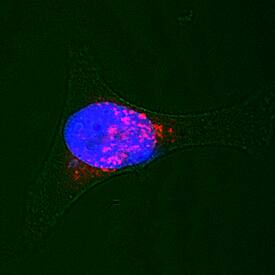
Syntaxin 7 was detected in immersion fixed HeLa human cervical epithelial carcinoma cell line using Sheep Anti-Human/Mouse/Rat Syntaxin 7 Antigen Affinity-purified Polyclonal Antibody (Catalog # AF5478) at 15 µg/mL for 3 hours at room temperature. Cells were stained using the NorthernLights™ 557-conjugated Anti-Sheep IgG Secondary Antibody (red; NL010) and counterstained with DAPI (blue). Specific staining was localized to lysosomes. View our protocol for Fluorescent ICC Staining of Cells on Coverslips.Applications for Human/Mouse/Rat Syntaxin 7 Antibody
Immunocytochemistry
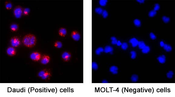
Sample: Immersion fixed HeLa Human Cervical Epithelial Carcinoma, Daudi Human Burkitt's Lymphoma (Positive) and MOLT‑4 Human Acute Lymphoblastic Leukemia (Negative) Cell Line Cells.
Western Blot
Sample: BJAB human Burkitt's lymphoma cell line, MCF-7 human breast cancer cell line, C2C12 mouse myoblast cell line, BaF3 mouse pro-B cell line, L1.2 mouse pro-B cell line, and Rat-2 rat embryonic fibroblast cell line
Reviewed Applications
Read 3 reviews rated 4 using AF5478 in the following applications:
Formulation, Preparation, and Storage
Purification
Reconstitution
Formulation
Shipping
Stability & Storage
- 12 months from date of receipt, -20 to -70 °C as supplied.
- 1 month, 2 to 8 °C under sterile conditions after reconstitution.
- 6 months, -20 to -70 °C under sterile conditions after reconstitution.
Background: Syntaxin 7
Syntaxin 7 (STX7) is a widely expressed protein embedded in endosomal and lysosomal membranes, and serves as a component of the SNARE complex. STX7 is involved in endocytic trafficking from early endosomes to late endosomes and lysosomes. This is in contrast to STX8, which is involved in clathrin-independent vesicular transport. Human STX7 is a type IV single-pass transmembrane protein (very short exoplasmic C-terminus) that is 261 amino acids (aa) in length. It contains a coiled-coil region (aa 47‑69), a t-SNARE domain (aa 165‑227) that is likely involved in protein-protein interactions, and a short, two amino acid, C-terminal luminal sequence. Over aa 21‑187, human STX7 shares 97% aa identity with mouse STX7.
Alternate Names
Gene Symbol
UniProt
Additional Syntaxin 7 Products
Product Documents for Human/Mouse/Rat Syntaxin 7 Antibody
Product Specific Notices for Human/Mouse/Rat Syntaxin 7 Antibody
For research use only


