Human/Mouse/Rat TC-PTP Antibody
R&D Systems, part of Bio-Techne | Catalog # MAB1930

Key Product Details
Validated by
Species Reactivity
Validated:
Cited:
Applications
Validated:
Cited:
Label
Antibody Source
Product Specifications
Immunogen
Pro2-Asn314
Accession # P17706
Specificity
Clonality
Host
Isotype
Scientific Data Images for Human/Mouse/Rat TC-PTP Antibody
Detection of Human/Mouse/Rat TC-PTP by Western Blot.
Western blot shows lysates of HepG2 human hepatocellular carcinoma cell line and TS1 mouse helper T cell line. PVDF membrane was probed with 0.5 µg/mL Mouse Anti-Human/Mouse/Rat TC-PTP Monoclonal Antibody (Catalog # MAB1930) followed by HRP-conjugated Anti-Mouse IgG Secondary Antibody (Catalog # HAF007). For additional reference, Recombinant Human PTP1B (Catalog # 1366-PT) and Recombinant Human TC-PTP aa 2-314 (Catalog # 1930-PT) (1 ng/lane) were included. A specific band for TC-PTP was detected at approximately 45 kDa (as indicated). This experiment was conducted under reducing conditions and using Immunoblot Buffer Group 1.Detection of Mouse TC-PTP/PTPN2 by Western Blot
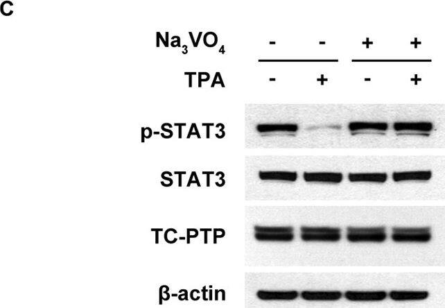
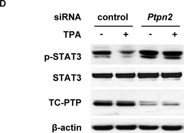
Effects of TC-PTP deficiency on STAT3 dephosphorylation in keratinocytes following TPA treatment.Total cell lysates were resolved by SDS-PAGE and immunoblotted with antibodies specific for STAT3 and phosphorylated STAT3 (p-STAT3). (A) 3PC keratinocytes were treated with 10, 20, 50 and 100 nM of TPA for 1 h. (B) 3PC keratinocytes were cultured in the presence of 5, 10, 20 and 50 μM Na3VO4 for 6 h. (C) 3PC keratinocyte were cultured with 50 μM of Na3VO4 for 2 h before treatment with 100 nM TPA. Cells were collected 6 h after TPA treatment. (D) 3PC keratinocytes were transfected with siRNA specific for Ptpn2. Cells were collected 1 h after TPA (100 nM) treatment. (E) 3PC keratinocytes were treated with 20, 50 and 100 nM of TPA for 1 h. Cells were collected at the indicated time after TPA treatment. (F–G) Control (scrambled) and TC-PTP-knockdown keratinocytes were treated with TPA (20 nM) for 1 h. Following TPA treatment, cells were collected at the indicated time. (G) *At 3 h of incubation, control and TC-PTP-knockdown keratinocytes were re-treated with TPA for 1 h. Cells were then collected 4 h after second TPA treatment. Image collected and cropped by CiteAb from the following publication (https://pubmed.ncbi.nlm.nih.gov/28322331), licensed under a CC-BY license. Not internally tested by R&D Systems.Detection of Mouse TC-PTP/PTPN2 by Western Blot
Effects of TC-PTP deficiency on STAT3 dephosphorylation in keratinocytes following TPA treatment.Total cell lysates were resolved by SDS-PAGE and immunoblotted with antibodies specific for STAT3 and phosphorylated STAT3 (p-STAT3). (A) 3PC keratinocytes were treated with 10, 20, 50 and 100 nM of TPA for 1 h. (B) 3PC keratinocytes were cultured in the presence of 5, 10, 20 and 50 μM Na3VO4 for 6 h. (C) 3PC keratinocyte were cultured with 50 μM of Na3VO4 for 2 h before treatment with 100 nM TPA. Cells were collected 6 h after TPA treatment. (D) 3PC keratinocytes were transfected with siRNA specific for Ptpn2. Cells were collected 1 h after TPA (100 nM) treatment. (E) 3PC keratinocytes were treated with 20, 50 and 100 nM of TPA for 1 h. Cells were collected at the indicated time after TPA treatment. (F–G) Control (scrambled) and TC-PTP-knockdown keratinocytes were treated with TPA (20 nM) for 1 h. Following TPA treatment, cells were collected at the indicated time. (G) *At 3 h of incubation, control and TC-PTP-knockdown keratinocytes were re-treated with TPA for 1 h. Cells were then collected 4 h after second TPA treatment. Image collected and cropped by CiteAb from the following publication (https://pubmed.ncbi.nlm.nih.gov/28322331), licensed under a CC-BY license. Not internally tested by R&D Systems.Applications for Human/Mouse/Rat TC-PTP Antibody
Western Blot
Sample: HepG2 human hepatocellular carcinoma cell line and TS1 mouse helper T cell line
Formulation, Preparation, and Storage
Purification
Reconstitution
Formulation
Shipping
Stability & Storage
- 12 months from date of receipt, -20 to -70 °C as supplied.
- 1 month, 2 to 8 °C under sterile conditions after reconstitution.
- 6 months, -20 to -70 °C under sterile conditions after reconstitution.
Background: TC-PTP
T-cell protein tyrosine phosphatase (TC-PTP), also known as PTPT and PTPN2, is an enzyme that removes phosphate groups covalently attached to tyrosine residues in proteins. This enzyme has two C-terminal end splice variants with distinctly different subcellular localizations. The shorter 45 kilodalton isoform is exclusively nuclear in resting cells, but redistrubutes to the cytosol upon stimulation with growth factors (1) and cellular stress (2). The longer 48 kilodalton isoform is exclusively found in the endoplasmic reticulum (3) and seems to have distinctly different physiologic substrates from the smaller isoform (1, 4). Although found in many cell types and tissues, TC-PTP is particularly prominent in hemopoietic cell types (5, 6). Knockout mice lacking TC-PTP are born viable but die 3 to 5 weeks after birth of erythropoietic and lymphopoietic deficits (7), indicating a critical role for TC-PTP in bone marrow maturation. TC-PTP will dephosphorylate a wide range of phosphoproteins, such as p52Shc (6) and receptors for EGF (1), Insulin (8) and growth hormone (6). The recombinant protein lacks the C-terminal 100 amino acids that determine intracellular localization but is fully active (9).
References
- Tiganis, T. et al. (1999) J. Biol. Chem. 274:27768.
- Lam, M.H. et al. (2001) J. Biol. Chem. 276:37700.
- Lorenzen, J.A. et al. (1995) J. Cell Biol. 131:631.
- Tiganis, T. et al. (1998) Mol. Cell. Biol. 18:1622.
- Cool, D.E. et al. (1989) Proc. Natl. Acad. Sci. USA 86:5257.
- Pasquali, C. et al. (2003) Mol. Endocrinol. 17:2228.
- You-Ten, K.E. et al. (1997) J. Exp. Med. 186:683.
- Galic, S. et al. (2003) Mol. Cell. Biol. 23:2096.
- Cool, D.E. et al. (1990) Proc. Natl. Acad. Sci. USA 87:7280.
Long Name
Alternate Names
Gene Symbol
UniProt
Additional TC-PTP Products
Product Documents for Human/Mouse/Rat TC-PTP Antibody
Product Specific Notices for Human/Mouse/Rat TC-PTP Antibody
For research use only


