Human/Mouse RBP4/Retinol-Binding Protein 4 Antibody
R&D Systems, part of Bio-Techne | Catalog # MAB34761


Key Product Details
Species Reactivity
Validated:
Cited:
Applications
Validated:
Cited:
Label
Antibody Source
Product Specifications
Immunogen
Met1-Leu201
Accession # NP_035385
Specificity
Clonality
Host
Isotype
Scientific Data Images for Human/Mouse RBP4/Retinol-Binding Protein 4 Antibody
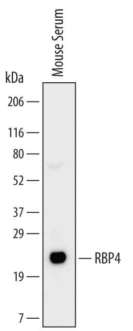
Detection of Mouse RBP4/Retinol‑Binding Protein 4 by Western Blot.
Western blot shows lysates of mouse serum. PVDF Membrane was probed with 2 µg/mL of Rat Anti-Human/Mouse RBP4/Retinol-Binding Protein 4 Monoclonal Antibody (Catalog # MAB34761) followed by HRP-conjugated Anti-Rat IgG Secondary Antibody (Catalog # HAF005). A specific band was detected for RBP4/Retinol-Binding Protein 4 at approximately 22 kDa (as indicated). This experiment was conducted under reducing conditions and using Immunoblot Buffer Group 1.Detection of Human RBP4/Retinol-Binding Protein 4 by Western Blot.
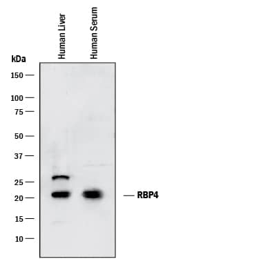
Western blot shows lysates of human liver tissue and human serum. PVDF membrane was probed with 0.05 µg/mL of Rat Anti-Human/Mouse RBP4/Retinol-Binding Protein 4 Monoclonal Antibody (Catalog # MAB34761) followed by HRP-conjugated Anti-Rat IgG Secondary Antibody (Catalog # HAF005). A specific band was detected for RBP4/Retinol-Binding Protein 4 at approximately 22 kDa (as indicated). This experiment was conducted under reducing conditions and using Immunoblot Buffer Group 1.Detection of RBP4/Retinol-Binding Protein 4 in Mouse Splenocytes by Flow Cytometry.
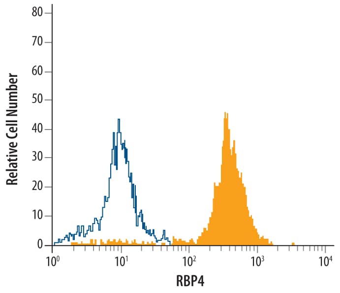
Mouse splenocytes were stained with Rat Anti-Human/Mouse RBP4/Retinol-Binding Protein 4 Monoclonal Antibody (Catalog # MAB34761, filled histogram) or isotype control antibody (Catalog # MAB006, open histogram), followed by Allophycocyanin-conjugated Anti-Rat IgG F(ab')2Secondary Antibody (Catalog # F0113).Applications for Human/Mouse RBP4/Retinol-Binding Protein 4 Antibody
CyTOF-ready
Flow Cytometry
Sample: Mouse splenocytes
Western Blot
Sample: Mouse serum, human liver tissue, and human serum
Formulation, Preparation, and Storage
Purification
Reconstitution
Formulation
Shipping
Stability & Storage
- 12 months from date of receipt, -20 to -70 °C as supplied.
- 1 month, 2 to 8 °C under sterile conditions after reconstitution.
- 6 months, -20 to -70 °C under sterile conditions after reconstitution.
Background: RBP4/Retinol-Binding Protein 4
Retinol (also known as vitamin A) is unstable and insoluble in the aqueous solution. However, retinol becomes quite stable and soluble in plasma due to its tight interaction with Retinol-binding Protein 4 (RBP4), also known as Plasma Retinol-binding Protein (1‑3). A prototypic member of the lipocalin superfamily, RBP4 has a beta‑barrel structure with a well-defined cavity. It is secreted from the liver, a process requiring the availability of retinol. RBP4 delivers retinol from the liver to the peripheral tissues. In plasma, the RBP4-retinol complex interacts with transthyretin (TTR), also known as thyroxine-binding protein and prealbumin. The retinol-RBP4-TTR complex prevents the loss of RBP4 by filtration through the kidney and increases the stability of the retinol-RBP4 complex. Defects in RBP4 cause retinol-binding protein deficiency, which affects night vision. Serum RBP4 levels are elevated in insulin-resistant mice and humans with obesity and type 2 diabetes, implying that RBP4, an adipocyte-derived signal, may be a biomarker and a drug target for the two diseases. The amino acid sequence of mouse RBP4 is 99%, 86%, 83% and 75% identical to that of rat, human/chimpanzee, dog and chicken.
References
- Zanotti, G. and R. Berni (2004) Vitamins and Hormones 69:271.
- Newcomer, M.E. and D.E. Ong (2000) Biochim. Biophys. Acta 1482:57.
- Yang, Q. et al. (2005) Nature 436:356.
Alternate Names
Gene Symbol
UniProt
Additional RBP4/Retinol-Binding Protein 4 Products
Product Documents for Human/Mouse RBP4/Retinol-Binding Protein 4 Antibody
Product Specific Notices for Human/Mouse RBP4/Retinol-Binding Protein 4 Antibody
For research use only

