Human/Mouse SOX2 PE-conjugated Antibody
R&D Systems, part of Bio-Techne | Catalog # IC2018P


Conjugate
Catalog #
Key Product Details
Species Reactivity
Validated:
Human, Mouse
Cited:
Human, Primate - Macaca fascicularis (Crab-eating Monkey or Cynomolgus Macaque)
Applications
Validated:
Intracellular Staining by Flow Cytometry
Cited:
Flow Cytometry
Label
Phycoerythrin (Excitation = 488 nm, Emission = 565-605 nm)
Antibody Source
Monoclonal Mouse IgG2A Clone # 245610
Product Specifications
Immunogen
E. coli-derived recombinant human SOX2
Gly135-Met317
Accession # P48431
Gly135-Met317
Accession # P48431
Specificity
Detects human and mouse SOX2 in Western blots.
Clonality
Monoclonal
Host
Mouse
Isotype
IgG2A
Scientific Data Images for Human/Mouse SOX2 PE-conjugated Antibody
Detection of SOX2 in NTera-2 Human Cell Line by Flow Cytometry.
NTera-2 human testicular embryonic carcinoma cell line was stained with Mouse Anti-Human/Mouse SOX2 PE-conjugated Monoclonal Antibody (Catalog # IC2018P, filled histogram) or isotype control antibody (Catalog # IC003P, open histogram). To facilitate intracellular staining, cells were fixed with Flow Cytometry Fixation Buffer (Catalog # FC004) and permeabilized with Flow Cytometry Permeabilization/Wash Buffer I (Catalog # FC005). View our protocol for Staining Intracellular Molecules.Detection of Human SOX2 by Flow Cytometry
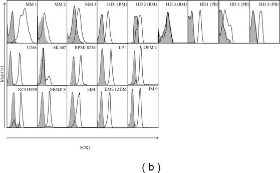
(a) RT-PCR analysis of SOX2 expression normalized to GAPDH in human tissues. BM from MM patients (n = 25), healthy donors (n = 15), myeloma cell lines (n = 10), and 20 human tissues (n = 1) was screened for SOX2 expression. Aqua dest. and non-reverse-transcribed mRNA were used as negative controls. 20 organs were tested for the presence of contaminating DNA. The resulting copy numbers (reverse-transcriptase-free) were normalized to GAPDH copy number of the respective tissue (cDNA). The mean value of all reverse-transcriptase-free results was calculated and included as the reverse-transcriptase-free (RT-free) condition. (b) FACS analysis of three MM patients' BM, three BM of healthy donors, and three peripheral blood samples of healthy donors for SOX2 expression in gated CD138+ plasma cells. One BM sample (3) was found negative for SO2 protein expression. SOX2 expression was also found in 10 different myeloma cell lines. Isotype antibodies served as negative control for SOX2 expression. (c) Correlation analysis of SOX2 expression and percentage of plasma cells in the BM of MM patients. No significant association between SOX2 expression and the amount of plasma cells was found (P = 0.6018, r2 = 0.03556). HD: healthy donor; MM: multiple myeloma; BM: bone marrow; PB: peripheral blood. Image collected and cropped by CiteAb from the following publication (https://pubmed.ncbi.nlm.nih.gov/22190969), licensed under a CC-BY license. Not internally tested by R&D Systems.Detection of Monkey SOX2 by Flow Cytometry
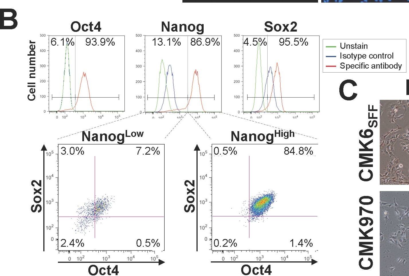
Pluripotency of Monkey ESCs grown under the MT-fCFA culture condition.A. Immunocytochemical analyses show that CMK6SFF cells (P33) have a characteristic expression pattern of typical pluripotency factors, Nanog, Oct4, and Sox2 as well as that of cell surface markers, SSEA-4, TRA-1-60, and TRA-1-81, indicating their undifferentiated and pluripotent state. Scale bar = 100 µm. B. Flow cytometric analysis of Nanog, Oct4, and Sox2 co-expressing CMK6SFF cells (P35) under the MT-fCFA culture condition. Cells were co-stained with Alexa Fluor 647-conjugated anti-Nanog, Alexa Fluor 488-conjugated anti-Oct4, and PE-conjugated anti-Sox2 or the corresponding isotype controls. C. CMK6SFF (P37) and CMK970 (P31) cells are positive for alkaline phosphatase activity. ALP, alkaline phosphatase. Scale bar = 100 µm. Image collected and cropped by CiteAb from the following publication (https://dx.plos.org/10.1371/journal.pone.0088346), licensed under a CC-BY license. Not internally tested by R&D Systems.Applications for Human/Mouse SOX2 PE-conjugated Antibody
Application
Recommended Usage
Intracellular Staining by Flow Cytometry
10 µL/106 cells
Sample: NTera‑2 human testicular embryonic carcinoma cell line fixed with Flow Cytometry Fixation Buffer (Catalog # FC004) and permeabilized with Flow Cytometry Permeabilization/Wash Buffer I (Catalog # FC005)
Sample: NTera‑2 human testicular embryonic carcinoma cell line fixed with Flow Cytometry Fixation Buffer (Catalog # FC004) and permeabilized with Flow Cytometry Permeabilization/Wash Buffer I (Catalog # FC005)
Formulation, Preparation, and Storage
Purification
Protein A or G purified from hybridoma culture supernatant
Formulation
Supplied in a saline solution containing BSA and Sodium Azide.
Shipping
The product is shipped with polar packs. Upon receipt, store it immediately at the temperature recommended below.
Stability & Storage
Protect from light. Do not freeze.
- 12 months from date of receipt, 2 to 8 °C as supplied.
Background: SOX2
References
- Graham, V. et al. (2003) Neuron 39:749.
- Avilion, A.A. et al. (2003) Genes Dev. 17:126.
- Kishi, M. et al. (2000) Development 127:791.
- Yuan, H. et al. (1995) Genes Dev. 9:2635.
- Uwanogho, D. et al. (1995) Mech. Dev. 49:23.
- Stevanovic, M. (2003) Mol. Biol. Rep. 30:127.
Long Name
Transcription Factor SOX2
Alternate Names
ANOP3, MCOPS3, MGC2413, SRY (sex determining region Y)-box 2, SRY-related HMG-box gene 2, transcription factor SOX2, transcription factor SOX-2
Entrez Gene IDs
6657 (Human)
Gene Symbol
SOX2
UniProt
Additional SOX2 Products
Product Documents for Human/Mouse SOX2 PE-conjugated Antibody
Product Specific Notices for Human/Mouse SOX2 PE-conjugated Antibody
For research use only
Loading...
Loading...
Loading...
Loading...
Loading...
Loading...

