Human/Mouse TOP2B Antibody
R&D Systems, part of Bio-Techne | Catalog # MAB6348

Key Product Details
Species Reactivity
Validated:
Cited:
Applications
Validated:
Cited:
Label
Antibody Source
Product Specifications
Immunogen
Phe1192-Asp1621
Accession # Q02880
Specificity
Clonality
Host
Isotype
Scientific Data Images for Human/Mouse TOP2B Antibody
Detection of Human and Mouse TOP2B by Western Blot.
Western blot shows lysates of HeLa human cervical epithelial carcinoma cell line, Jurkat human acute T cell leukemia cell line, MOLT-4 human acute lymphoblastic leukemia cell line, K562 human chronic myelogenous leukemia cell line, and CH-1 mouse B cell lymphoma cell line. PVDF Membrane was probed with 1 µg/mL of Mouse Anti-Human/Mouse TOP2B Monoclonal Antibody (Catalog # MAB6348) followed by HRP-conjugated Anti-Mouse IgG Secondary Antibody (Catalog # HAF007). A specific band was detected for TOP2B at approximately 185 kDa (as indicated). This experiment was conducted under reducing conditions and using Immunoblot Buffer Group 1.TOP2B in HeLa Human Cell Line.
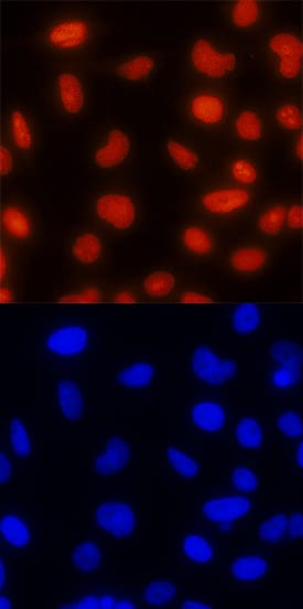
TOP2B was detected in immersion fixed HeLa human cervical epithelial carcinoma cell line using Mouse Anti-Human/Mouse TOP2B Monoclonal Antibody (Catalog # MAB6348) at 25 µg/mL for 3 hours at room temperature. Cells were stained using the NorthernLights™ 557-conjugated Anti-Mouse IgG Secondary Antibody (red, upper panel; Catalog # NL007) and counterstained with DAPI (blue, lower panel). Specific staining was localized to nuclei. View our protocol for Fluorescent ICC Staining of Cells on Coverslips.Applications for Human/Mouse TOP2B Antibody
Immunocytochemistry
Sample: Immersion fixed HeLa human cervical epithelial carcinoma cell line
Western Blot
Sample: HeLa human cervical epithelial carcinoma cell line, Jurkat human acute T cell leukemia cell line, MOLT‑4 human acute lymphoblastic leukemia cell line, K562 human chronic myelogenous leukemia cell line, and CH‑1 mouse B cell lymphoma cell line
Formulation, Preparation, and Storage
Purification
Reconstitution
Formulation
Shipping
Stability & Storage
- 12 months from date of receipt, -20 to -70 °C as supplied.
- 1 month, 2 to 8 °C under sterile conditions after reconstitution.
- 6 months, -20 to -70 °C under sterile conditions after reconstitution.
Background: TOP2B
TOP2B (DNA Topoisomerase II beta) is a 180-185 kDa member of the type IIA subfamily, topoisomerase family of molecules. It is ubiquitously expressed, and represents the larger of two known topoisomerases (the 2A/a-form being 170 kDa in size). TOP2A is essential for cell division, while TOP2B is active postmitotically. In an ATP-dependent manner, homodimeric TOP2B relives torsional stress created in DNA during transcription or as a consequence of replication. TOP2B first induces cleavage of double-stranded DNA, creating a space that allows for the physical repositioning of chromatin and a reduction in tension. This is followed by closure and ligation of the cleaved ends to recreate the original DNA structure. Human TOP2B is 1626 amino acids (aa) in length. It contains an ATPase domain (aa 101-201), an NES (aa 1034-1044), more that 30 Ser/Thr phosphorylation sites and a C-terminal DTHCT region (aa 1508-1611). There is one splice variant that shows a deletion of aa 24-28. Over aa 1187-1621, human TOP2B shares 91% aa identity with mouse TOP2B.
Long Name
Alternate Names
Gene Symbol
UniProt
Additional TOP2B Products
Product Documents for Human/Mouse TOP2B Antibody
Product Specific Notices for Human/Mouse TOP2B Antibody
For research use only

