Human Myocilin Antibody
R&D Systems, part of Bio-Techne | Catalog # MAB3446

Key Product Details
Species Reactivity
Validated:
Human
Cited:
Human, Tree Shrew
Applications
Validated:
Immunohistochemistry
Cited:
Immunohistochemistry, Immunoprecipitation, Western Blot
Label
Unconjugated
Antibody Source
Monoclonal Mouse IgG2B Clone # 297817
Product Specifications
Immunogen
Mouse myeloma cell line NS0-derived recombinant human Myocilin
Arg33-Met504
Accession # Q99972
Arg33-Met504
Accession # Q99972
Specificity
Detects human Myocilin in direct ELISAs. In direct ELISAs, 75% cross-reactivity with recombinant mouse Myocilin is observed.
Clonality
Monoclonal
Host
Mouse
Isotype
IgG2B
Scientific Data Images for Human Myocilin Antibody
Myocilin in Human Skeletal Muscle.
Myocilin was detected in immersion fixed paraffin-embedded sections of Human Skeletal Muscle using Mouse Anti-Human Myocilin Monoclonal Antibody (Catalog # MAB3446) at 5 µg/mL for 1 hour at room temperature followed by incubation with the Anti-Goat IgG VisUCyte™ HRP Polymer Antibody (Catalog # VC004). Before incubation with the primary antibody, tissue was subjected to heat-induced epitope retrieval using VisUCyte Antigen Retrieval Reagent-Basic (Catalog # VCTS021). Tissue was stained using DAB (brown) and counterstained with hematoxylin (blue). Specific staining was localized to sarcoplasm and sarcolemma in skeletal muscle cells. View our protocol for IHC Staining with VisUCyte HRP Polymer Detection Reagents.Detection of Mouse Myocilin by Western Blot
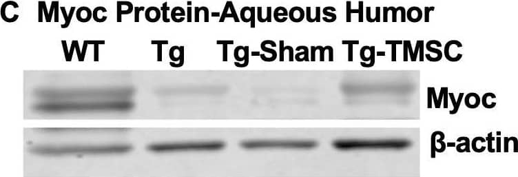
TMSCs reduce the Myoc retention in the TM tissue, promote the Myoc secretion into the aqueous humor, and reverse the ECM expression in the Tg-MyocY437H mice.(A) Immunofluorescent staining shows accumulated Myoc in the TM, iris, and ciliary body of the Tg and Tg-sham mice. TMSC transplantation alleviated the aggregation of Myoc in the TM, similar to the WT mice. Scale bars, 50 mm. Western blotting results show: (B) The representative bands of Myoc expression in the mouse limbal tissue and the relative Myoc protein levels with b-actin as internal control (n=5). (C) The representative bands of Myoc expression in the mouse aqueous humor and the relative Myoc protein levels with b-actin as internal control (n=5). (D) The representative bands of the expression of ECM components fibronectin (FN), collagen IV, and elastin in the limbal tissue and the relative ECM protein levels with b-actin as internal control (n=4-6). Data are presented as mean ± SD. One-way ANOVA (B,C) or two-way ANOVA (D) followed by Tukey’s multiple comparisons test. C: cornea, SC: Schlemm’s canal, TM: trabecular meshwork.Figure 5—source data 1.Relative myocilin protein levels in the corneal limbus for Figure 5B.Relative myocilin protein levels in the aqueous humor of each eye for Figure 5C; Relative extracellular mactrix protein levels in the corneal limbus for Figure 5D.Relative myocilin protein levels in the corneal limbus for Figure 5B.Relative myocilin protein levels in the aqueous humor of each eye for Figure 5C; Relative extracellular mactrix protein levels in the corneal limbus for Figure 5D. Image collected and cropped by CiteAb from the following open publication (https://pubmed.ncbi.nlm.nih.gov/33506763), licensed under a CC-BY license. Not internally tested by R&D Systems.Detection of Mouse Myocilin by Western Blot
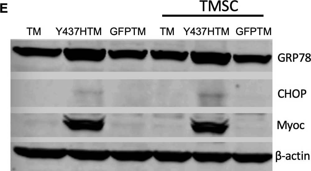
TMSCs could not reverse ER stress and stimulate proliferation of Myoc mutant TM cells in vitro.(A) The TM cells were transduced with recombinant lentivirus encoding GFP and Myoc Y437H mutation. The transduced GFP+ cells were sorted by Flow cytometry and the cultured sorted TM cells were almost 100% with GFP (green) in the cytoplasm. Scale Bars, 100 µm. (B) Transduced TM cells with Myoc Y437H mutation expressed high Myoc by western blotting and TM cells had increased Myoc expression after 5-day Dex treatment (TM2, TM3). TM1 did not have increased Myoc expression after Dex treatment so TM1 cells were discarded. (C) Schematic illustration shows co-culturing of TMSCs with TM cells for detection of TM cell changes. (D) Flow cytometry analysis of EdU incorporation shows neither co-culture nor direct contact with TMSCs for 4 days would affect TM cell proliferation (n=3-5). (E) Representative western blotting bands show the levels of ER stress markers and Myoc in the TM cells with or without TMSC co-culturing. (F) Relative protein levels with b-actin as internal control (n=3). Data are presented as mean ± SD. One-way ANOVA (D) or two-way ANOVA (F) followed by Tukey’s multiple comparisons test.Figure 7—source data 1.Percentage of BrdU positive TM cells in different culture conditions for Figure 7D.Relative protein levels by WB in the TM cells with different culture conditions for Figure 7F.Percentage of BrdU positive TM cells in different culture conditions for Figure 7D.Relative protein levels by WB in the TM cells with different culture conditions for Figure 7F.Structure of lentiviral packaging plasmid: pLentiCMV-Y437H-IRES-GFP. Image collected and cropped by CiteAb from the following open publication (https://pubmed.ncbi.nlm.nih.gov/33506763), licensed under a CC-BY license. Not internally tested by R&D Systems.Applications for Human Myocilin Antibody
Application
Recommended Usage
Immunohistochemistry
5-25 µg/mL
Sample: Immersion fixed paraffin-embedded sections of Human Skeletal Muscle
Sample: Immersion fixed paraffin-embedded sections of Human Skeletal Muscle
Reviewed Applications
Read 1 review rated 5 using MAB3446 in the following applications:
Formulation, Preparation, and Storage
Purification
Protein A or G purified from hybridoma culture supernatant
Reconstitution
Reconstitute at 0.5 mg/mL in sterile PBS. For liquid material, refer to CoA for concentration.
Formulation
Lyophilized from a 0.2 μm filtered solution in PBS with Trehalose. *Small pack size (SP) is supplied either lyophilized or as a 0.2 µm filtered solution in PBS.
Shipping
Lyophilized product is shipped at ambient temperature. Liquid small pack size (-SP) is shipped with polar packs. Upon receipt, store immediately at the temperature recommended below.
Stability & Storage
Use a manual defrost freezer and avoid repeated freeze-thaw cycles.
- 12 months from date of receipt, -20 to -70 °C as supplied.
- 1 month, 2 to 8 °C under sterile conditions after reconstitution.
- 6 months, -20 to -70 °C under sterile conditions after reconstitution.
Background: Myocilin
Additional Myocilin Products
Product Documents for Human Myocilin Antibody
Product Specific Notices for Human Myocilin Antibody
For research use only
Loading...
Loading...
Loading...
Loading...


