Human N-Cadherin Antibody
R&D Systems, part of Bio-Techne | Catalog # MAB13881


Key Product Details
Species Reactivity
Validated:
Human
Cited:
Human
Applications
Validated:
CyTOF-ready, Flow Cytometry, Immunohistochemistry, Western Blot
Cited:
Immunoprecipitation, Western Blot
Label
Unconjugated
Antibody Source
Monoclonal Mouse IgM Clone # 691723
Product Specifications
Immunogen
Mouse myeloma cell line NS0-derived recombinant mouse N-Cadherin
Asp160-Ala724
Accession # P19022.4
Asp160-Ala724
Accession # P19022.4
Specificity
Detects mouse N-Cadherin in direct ELISAs and Western blots. In direct ELISAs approximately 50% cross-reactivity
with recombinant mouse N-Cadherin is observed, and no cross-reactivity with recombinant
human (rh) E-Cadherin, rhP-Cadherin, rhVE-Cadherin, rhCadherin-4, -8, -11, -12,
or -13 is observed.
Clonality
Monoclonal
Host
Mouse
Isotype
IgM
Scientific Data Images for Human N-Cadherin Antibody
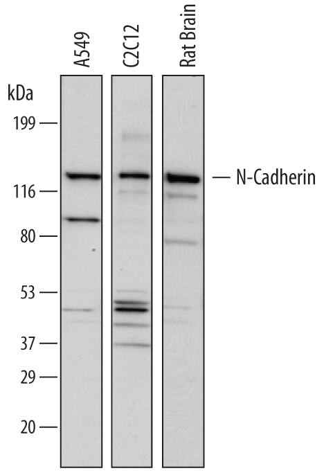
Detection of Human, Mouse, and Rat N-Cadherin by Western Blot.
Western blot shows lysates of A549 human lung carcinoma cell line, C2C12 mouse myoblast cell line, and rat brain tissue. PVDF membrane was probed with 1 µg/mL of Mouse Anti-Human N-Cadherin Monoclonal Antibody (Catalog # MAB13881) followed by HRP-conjugated Anti-Mouse IgG Secondary Antibody (Catalog # HAF007). A specific band was detected for N-Cadherin at approximately 130 kDa (as indicated). This experiment was conducted under reducing conditions and using Immunoblot Buffer Group 1.Detection of N-Cadherin in HeLa Human Cell Line by Flow Cytometry.
HeLa human cervical epithelial carcinoma cell line was stained with Mouse Anti-Human N-Cadherin Monoclonal Antibody (Catalog # MAB13881, filled histogram) or isotype control antibody mouse IgM (open histogram), followed by Phycoerythrin-conjugated Anti-Mouse IgM Secondary Antibody (Catalog # F0116).N‑Cadherin in Human Brain.
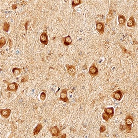
N-Cadherin was detected in immersion fixed paraffin-embedded sections of Alzheimer's human brain using Mouse Anti-Human N-Cadherin Monoclonal Antibody (Catalog # MAB13881) at 15 µg/mL overnight at 4 °C. Before incubation with the primary antibody, tissue was subjected to heat-induced epitope retrieval using Antigen Retrieval Reagent-Basic (Catalog # CTS013). Tissue was stained using the Anti-Mouse HRP-DAB Cell & Tissue Staining Kit (brown; Catalog # CTS002) and counterstained with hematoxylin (blue). Specific staining was localized to cytoplasm of neurons. View our protocol for Chromogenic IHC Staining of Paraffin-embedded Tissue Sections.Applications for Human N-Cadherin Antibody
Application
Recommended Usage
CyTOF-ready
Ready to be labeled using established conjugation methods. No BSA or other carrier proteins that could interfere with conjugation.
Flow Cytometry
2.5 µg/106 cells
Sample: HeLa human cervical epithelial carcinoma cell line
Sample: HeLa human cervical epithelial carcinoma cell line
Immunohistochemistry
8-25 µg/mL
Sample: Immersion fixed paraffin-embedded sections of human Alzheimer's brain
Sample: Immersion fixed paraffin-embedded sections of human Alzheimer's brain
Western Blot
1 µg/mL
Sample: A549 human lung carcinoma cell line, C2C12 mouse myoblast cell line, and rat brain tissue
Sample: A549 human lung carcinoma cell line, C2C12 mouse myoblast cell line, and rat brain tissue
Reviewed Applications
Read 1 review rated 3 using MAB13881 in the following applications:
Formulation, Preparation, and Storage
Purification
Protein A or G purified from hybridoma culture supernatant
Reconstitution
Sterile PBS to a final concentration of 0.5 mg/mL. For liquid material, refer to CoA for concentration.
Formulation
Lyophilized from a 0.2 μm filtered solution in PBS with Trehalose. *Small pack size (SP) is supplied either lyophilized or as a 0.2 µm filtered solution in PBS.
Shipping
Lyophilized product is shipped at ambient temperature. Liquid small pack size (-SP) is shipped with polar packs. Upon receipt, store immediately at the temperature recommended below.
Stability & Storage
Use a manual defrost freezer and avoid repeated freeze-thaw cycles.
- 12 months from date of receipt, -20 to -70 °C as supplied.
- 1 month, 2 to 8 °C under sterile conditions after reconstitution.
- 6 months, -20 to -70 °C under sterile conditions after reconstitution.
Background: N-Cadherin
References
- Pokutta, S. and W.I. Weis (2007) Annu. Rev. Cell Dev. Biol. 23:237.
- Gumbiner, B.M. (2005) Nat. Rev. Mol. Cell Biol. 6:622.
- Reid, R.A. and J.J. Hemperly (1990) Nucleic Acids Res. 18:5896.
- Wanner, I.B. and P.M. Wood (2002) J. Neurosci. 22:4066.
- Benson, D.L. and H. Tanaka (1998) J. Neurosci. 18:6892.
- Saglietti, L. et al. (2007) Neuron 54:461.
- Reiss, K. et al. (2005) EMBO J. 24:742.
- Malinverno, M. et al. (2010) J. Neurosci. 30:16343.
- Jang, Y.-N. et al. (2009) J. Neurosci. 29:5974.
- Uemura, K. et al. (2006) Neurosci. Lett. 402:278.
- Hartland, S.N. et al. (2009) Liver Int. 29:966.
- Williams, H. et al. (2010) Cardiovasc. Res. 87:137.
- Dwivedi, A. et al. (2009) Cardiovasc. Res. 81:178.
- Maret, D. et al. (2010) Neoplasia 12:1066.
- Tanaka, H. et al. (2010) Nat. Med. 16:1414.
- Wein, F. et al. (2010) Stem Cell Res. 4:129.
Long Name
Neural Cadherin
Alternate Names
Cadherin-2, CD325, CDH2, NCadherin
Gene Symbol
CDH2
UniProt
Additional N-Cadherin Products
Product Documents for Human N-Cadherin Antibody
Product Specific Notices for Human N-Cadherin Antibody
For research use only
Loading...
Loading...
Loading...
Loading...
Loading...

