Human Nectin-4 Antibody
R&D Systems, part of Bio-Techne | Catalog # AF2659


Key Product Details
Validated by
Knockout/Knockdown
Species Reactivity
Validated:
Human
Cited:
Human, Mouse, Porcine, Canine, Hamster, Primate - Chlorocebus aethiops (African Green Monkey), Primate - Macaca fascicularis (Crab-eating Monkey or Cynomolgus Macaque), Xenograft
Applications
Validated:
CyTOF-ready, Flow Cytometry, Immunohistochemistry, Western Blot
Cited:
Flow Cytometry, Functional Assay, Immunocytochemistry, Immunohistochemistry, Immunohistochemistry-Paraffin, Neutralization, Westen Blot, Western Blot
Label
Unconjugated
Antibody Source
Polyclonal Goat IgG
Product Specifications
Immunogen
Mouse myeloma cell line NS0-derived recombinant human Nectin-4
Gly27-Val351
Accession # Q96NY8
Gly27-Val351
Accession # Q96NY8
Specificity
Detects human Nectin-4 in direct ELISAs and Western blots. In direct ELISAs, approximately 75% cross‑reactivity with recombinant mouse Nectin-4 is observed.
Clonality
Polyclonal
Host
Goat
Isotype
IgG
Scientific Data Images for Human Nectin-4 Antibody
Nectin-4 in Human Placenta.
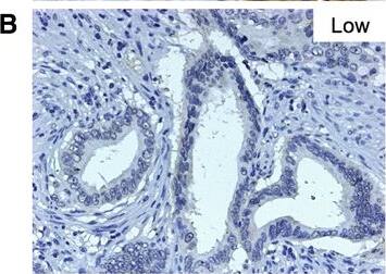
Nectin-4 was detected in immersion fixed paraffin-embedded sections of human placenta using 10 µg/mL Goat Anti-Human Nectin-4 Antigen Affinity-purified Polyclonal Antibody (Catalog # AF2659) overnight at 4 °C. Before incubation with the primary antibody tissue was subjected to heat-induced epitope retrieval using Antigen Retrieval Reagent-Basic (CTS013). Tissue was stained with the Anti-Goat HRP-DAB Cell & Tissue Staining Kit (brown; CTS008) and counterstained with hematoxylin (blue). View our protocol for Chromogenic IHC Staining of Paraffin-embedded Tissue Sections.Detection of Human Nectin-4/PVRL4 by Western Blot
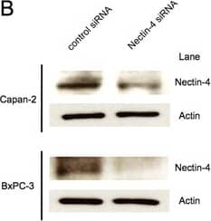
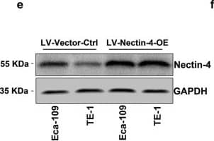
Inhibition of Nectin-4 expression by gene silencing decreases cell proliferation in pancreatic cancer cells. (A,B) Capan-2 and BxPC-3 cells were transfected with Nectin-4 siRNA or control RNA. The relative expression of Nectin-4 was significantly reduced in both cells when transfected with Nectin-4 siRNA for up to 72 hours as determined by quantitative real-time PCR and Western blot analysis (n = 4 of each group). (C) Cell proliferation was significantly inhibited by Nectin-4 gene silencing in both cells after 72 hours incubation as determined by MTS assay (n = 6 of each group). *P < 0.001 versus control siRNA (Student’s t test). Image collected and cropped by CiteAb from the following publication (https://jeccr.biomedcentral.com/articles/10.1186/s13046-015-0144-7), licensed under a CC-BY license. Not internally tested by R&D Systems.Detection of Nectin-4 in MCF-7 Human Cell Line by Flow Cytometry.
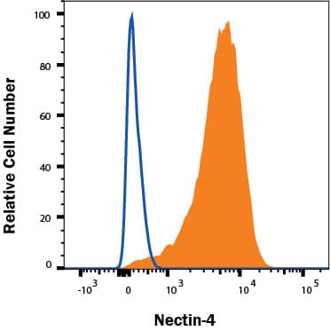
MCF-7 human breast adenocarcinoma cell line was stained with Goat Anti-Human Nectin-4 Antigen Affinity-purified Polyclonal Antibody (Catalog # AF2659, filled histogram) or control antibody (AB-108-C, open histogram), followed by PE-conjugated Anti-Goat IgG Secondary Antibody (F0107).Applications for Human Nectin-4 Antibody
Application
Recommended Usage
CyTOF-ready
Ready to be labeled using established conjugation methods. No BSA or other carrier proteins that could interfere with conjugation.
Flow Cytometry
0.25 µg/106 cells
Sample: MCF-7 human breast cancer cell line
Sample: MCF-7 human breast cancer cell line
Immunohistochemistry
5-15 µg/mL
Sample: Immersion fixed paraffin-embedded sections of human placenta subjected to Antigen Retrieval Reagent-Basic (Catalog # CTS013)
Sample: Immersion fixed paraffin-embedded sections of human placenta subjected to Antigen Retrieval Reagent-Basic (Catalog # CTS013)
Western Blot
0.1 µg/mL
Sample: Recombinant Human Nectin‑4 (Catalog # 2659-N4)
Sample: Recombinant Human Nectin‑4 (Catalog # 2659-N4)
Formulation, Preparation, and Storage
Purification
Antigen Affinity-purified
Reconstitution
Reconstitute at 0.2 mg/mL in sterile PBS. For liquid material, refer to CoA for concentration.
Formulation
Lyophilized from a 0.2 μm filtered solution in PBS with Trehalose. *Small pack size (SP) is supplied either lyophilized or as a 0.2 µm filtered solution in PBS.
Shipping
Lyophilized product is shipped at ambient temperature. Liquid small pack size (-SP) is shipped with polar packs. Upon receipt, store immediately at the temperature recommended below.
Stability & Storage
Use a manual defrost freezer and avoid repeated freeze-thaw cycles.
- 12 months from date of receipt, -20 to -70 °C as supplied.
- 1 month, 2 to 8 °C under sterile conditions after reconstitution.
- 6 months, -20 to -70 °C under sterile conditions after reconstitution.
Background: Nectin-4
Long Name
Poliovirus Receptor Related 4
Alternate Names
LNIR, Nectin4, PRR4, PVRL4
Gene Symbol
NECTIN4
UniProt
Additional Nectin-4 Products
Product Documents for Human Nectin-4 Antibody
Product Specific Notices for Human Nectin-4 Antibody
For research use only
Loading...
Loading...
Loading...
Loading...
Loading...
