Human Nodal Antibody
R&D Systems, part of Bio-Techne | Catalog # MAB3218

Key Product Details
Species Reactivity
Applications
Label
Antibody Source
Product Specifications
Immunogen
His238-Leu347
Accession # Q96S42
Specificity
Clonality
Host
Isotype
Scientific Data Images for Human Nodal Antibody
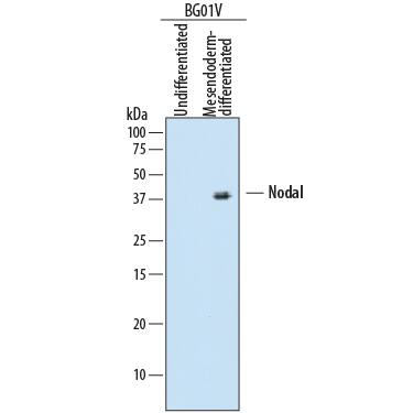
Detection of Human Nodal by Western Blot.
Western blot shows lysates of undifferentiated or mesendoderm-differentiated BG01V human embryonic stem cells. PVDF membrane was probed with 2 µg/mL of Mouse Anti-Human Nodal Monoclonal Antibody (Catalog # MAB3218) followed by HRP-conjugated Anti-Mouse IgG Secondary Antibody (Catalog # HAF007). A specific band was detected for Nodal at approximately 35-47 kDa (as indicated). This experiment was conducted under reducing conditions and using Immunoblot Buffer Group 1.Applications for Human Nodal Antibody
Western Blot
Sample: Undifferentiated or mesendoderm-differentiated BG01V human embryonic stem cells
Formulation, Preparation, and Storage
Purification
Reconstitution
Formulation
Shipping
Stability & Storage
- 12 months from date of receipt, -20 to -70 °C as supplied.
- 1 month, 2 to 8 °C under sterile conditions after reconstitution.
- 6 months, -20 to -70 °C under sterile conditions after reconstitution.
Background: Nodal
Nodal is a secreted protein that is a member of the Transforming Growth Factor-beta (TGF-beta) superfamily. Nodal was named for its localized expression in the mouse node during gastrulation, and is first detected in the early primitive streak during mesoderm formation. Expression of the Nodal gene occurs asymmetrically in the left, but not right, lateral plate during somitogenesis. Nodal proteins play crucial roles in mesoderm formation and both anterior-posterior and left-right axis formation during vertebrate development. Members of the Nodal gene family include mouse Nodal, chick cNR-1, frog Xnr1-4, and zebrafish cyclops. Biologically active Nodal is a disulfide-linked homodimer and contains all seven of the cysteine residues necessary for formation of the “cysteine knot” characteristic of TGF-beta -related molecules. Mouse Nodal is 34-39% homologous in the conserved region to other TGF-beta superfamily members. Nodal has been shown to signal through a mechanism related to the Activin pathway, and signaling is mediated through both Smad2 and 3. Nodal signaling utilizes type II Activin receptors, together with ALK4/ActRIB, or the type I receptor ALK7. Nodal interacts extracellularly with members of other protein families including Cerberus, Lefty, and EGF-CFC ligands such as Cripto. While the Cerberus and Lefty families act as Nodal antagonists, the EGF-CFC molecules act as co-receptors to facilitate Nodal signaling. The resulting concert of regulated Nodal activity allows for the precise control of mesoderm formation, neural patterning, and axis positioning and patterning during early vertebrate development.
References
- Kumar, A. et al. (2001) J. Biol. Chem. 276:656.
- Reissmann, E. et al. (2001) Genes & Dev. 15: 2010.
- Schier, A. and M. Shen (1999) Nature 403:385.
- Zhou, X. et al. (1993) Nature 361:543.
Alternate Names
Gene Symbol
UniProt
Additional Nodal Products
Product Documents for Human Nodal Antibody
Product Specific Notices for Human Nodal Antibody
For research use only
