Human OPA1 Antibody
R&D Systems, part of Bio-Techne | Catalog # MAB9506
Recombinant Monoclonal Antibody.

Key Product Details
Species Reactivity
Human
Applications
Simple Western, Western Blot
Label
Unconjugated
Antibody Source
Recombinant Monoclonal Rabbit IgG Clone # 1284B
Product Specifications
Immunogen
A synthetic peptide made to an internal region within
amino acid residues 500-600 of human OPA1
Accession # O60313
Accession # O60313
Specificity
Detects human, mouse, and rat OPA1 in Western blots.
Clonality
Monoclonal
Host
Rabbit
Isotype
IgG
Scientific Data Images for Human OPA1 Antibody
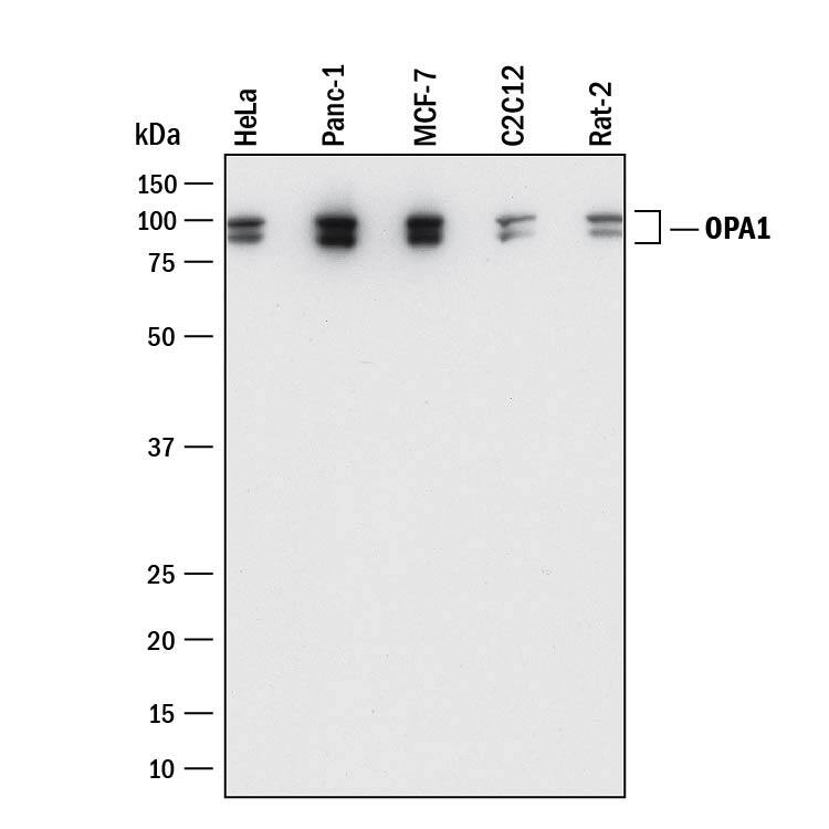
Detection of Human, Mouse, and Rat OPA1 by Western Blot.
Western blot shows lysates of HeLa human cervical epithelial carcinoma cell line, PANC-1 human pancreatic carcinoma cell line, MCF-7 human breast cancer cell line, C2C12 mouse myoblast cell line, and Rat-2 rat embryonic fibroblast cell line. PVDF membrane was probed with 0.2 µg/mL of Rabbit Anti-Human OPA1 Monoclonal Antibody (Catalog # MAB9506) followed by HRP-conjugated Anti-Rabbit IgG Secondary Antibody (Catalog # HAF008). Specific bands were detected for OPA1 at approximately 80-100 kDa (as indicated). This experiment was conducted under reducing conditions and using Immunoblot Buffer Group 1.Detection of Human OPA1 by Simple WesternTM.
Simple Western shows lysates of MDA-MB-231 human breast cancer cell line, loaded at 0.5 mg/ml. A specific band was detected for OPA1 at approximately 90-96 kDa (as indicated) using 20 µg/mL of Rabbit Anti-Human OPA1 Monoclonal Antibody (Catalog # MAB9506). This experiment was conducted under reducing conditions and using the 12-230kDa separation system.Applications for Human OPA1 Antibody
Application
Recommended Usage
Simple Western
20 µg/mL
Sample: MDA-MB-231 human breast cancer cell line
Sample: MDA-MB-231 human breast cancer cell line
Western Blot
0.2 µg/mL
Sample: HeLa human cervical epithelial carcinoma cell line, PANC-1 human pancreatic carcinoma cell line, MCF-7 human breast cancer cell line, C2C12 mouse myoblast cell line, and Rat-2 rat embryonic fibroblast cell line
Sample: HeLa human cervical epithelial carcinoma cell line, PANC-1 human pancreatic carcinoma cell line, MCF-7 human breast cancer cell line, C2C12 mouse myoblast cell line, and Rat-2 rat embryonic fibroblast cell line
Formulation, Preparation, and Storage
Purification
Protein A or G purified from cell culture supernatant
Reconstitution
Reconstitute at 0.5 mg/mL in sterile PBS. For liquid material, refer to CoA for concentration.
Formulation
Lyophilized from a 0.2 μm filtered solution in PBS with Trehalose. See Certificate of Analysis for details.
*Small pack size (-SP) is supplied either lyophilized or as a 0.2 µm filtered solution in PBS.
*Small pack size (-SP) is supplied either lyophilized or as a 0.2 µm filtered solution in PBS.
Shipping
Lyophilized product is shipped at ambient temperature. Liquid small pack size (-SP) is shipped with polar packs. Upon receipt, store immediately at the temperature recommended below.
Stability & Storage
Use a manual defrost freezer and avoid repeated freeze-thaw cycles.
- 12 months from date of receipt, -20 to -70 °C as supplied.
- 1 month, 2 to 8 °C under sterile conditions after reconstitution.
- 6 months, -20 to -70 °C under sterile conditions after reconstitution.
Background: OPA1
Long Name
Optic Atrophy Protein 1
Alternate Names
BERHS, EC 3.6.5.5, LargeG, lilr3, MGM1, MTDPS14, NPG, NTG
Gene Symbol
OPA1
UniProt
Additional OPA1 Products
Product Documents for Human OPA1 Antibody
Product Specific Notices for Human OPA1 Antibody
For research use only
Loading...
Loading...
Loading...
Loading...

