Human Osteopontin/OPN Antibody
R&D Systems, part of Bio-Techne | Catalog # MAB14334

Key Product Details
Species Reactivity
Applications
Label
Antibody Source
Product Specifications
Immunogen
Accession # NP_000573.1
Specificity
Clonality
Host
Isotype
Scientific Data Images for Human Osteopontin/OPN Antibody
Osteopontin/OPN in HepG2 Human Cell Line.
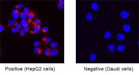
Osteopontin/OPN was detected in immersion fixed HepG2 human hepatocellular carcinoma cell line (positive) and Daudi human Burkitt's lymphoma cell line (negative control) using Rabbit Anti-Human Osteopontin/OPN Monoclonal Antibody (Catalog # MAB14334) at 3 µg/mL for 3 hours at room temperature. Cells were stained using the NorthernLights™ 557-conjugated Anti-Rabbit IgG Secondary Antibody (red; Catalog # NL004) and counterstained with DAPI (blue). Specific staining was localized to cytoplasm. View our protocol for Fluorescent ICC Staining of Cells on Coverslips.Osteopontin/OPN in Human Kidney.
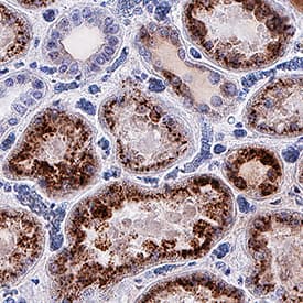
Osteopontin/OPN was detected in immersion fixed paraffin-embedded sections of human kidney using Rabbit Anti-Human Osteopontin/OPN Monoclonal Antibody (Catalog # MAB14334) at 3 µg/mL for 1 hour at room temperature followed by incubation with the Anti-Rabbit IgG VisUCyte™ HRP Polymer Antibody (Catalog # VC003). Before incubation with the primary antibody, tissue was subjected to heat-induced epitope retrieval using Antigen Retrieval Reagent-Basic (Catalog # CTS013). Tissue was stained using DAB (brown) and counterstained with hematoxylin (blue). Specific staining was localized to cytoplasm in convoluted tubules. View our protocol for IHC Staining with VisUCyte HRP Polymer Detection Reagents.Cell Adhesion Mediated by Osteopontin/OPN and Neutralization by Human Osteopontin/OPN Antibody.
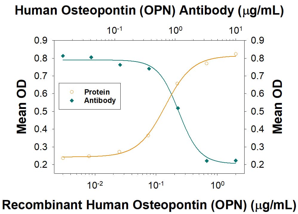
Recombinant Human Osteopontin/OPN (Catalog # 1433-OP), immobilized onto a microplate, supports the adhesion of the HEK293 human embryonic kidney cell line in a dose-dependent manner (orange line). Adhesion elicited by Recombinant Human Osteopontin/OPN (1 µg/mL) is neutralized (green line) by increasing concentrations of Rabbit Anti-Human Osteopontin/OPN Monoclonal Antibody (Catalog # MAB14334). The ND50 is typically 0.5-1.5 µg/mL.Applications for Human Osteopontin/OPN Antibody
Immunocytochemistry
Sample: Immersion fixed HepG2 human hepatocellular carcinoma cell line
Immunohistochemistry
Sample: Immersion fixed paraffin-embedded sections of human kidney
Neutralization
Formulation, Preparation, and Storage
Purification
Reconstitution
Formulation
Shipping
Stability & Storage
- 12 months from date of receipt, -20 to -70 °C as supplied.
- 1 month, 2 to 8 °C under sterile conditions after reconstitution.
- 6 months, -20 to -70 °C under sterile conditions after reconstitution.
Background: Osteopontin/OPN
Osteopontin (OPN, previously also referred to as transformation-associated secreted phosphoprotein, bone sialoprotein I, 2ar, 2B7, early T lymphocyte activation 1 protein, minopotin, calcium oxalate crystal growth inhibitor protein), is a secreted, highly acidic, calcium-binding, RGD-containing, phosphorylated glycoprotein originally isolated from bone matrix (1). Subsequently, OPN has been found in kidney, placenta, blood vessels and various tumor tissues. Many cell types (including macrophages, osteoclasts, activated T-cells, fibroblasts, epithelial cells, vascular smooth muscle cells, and natural killer cells) can express OPN in response to activation by cytokines, growth factors or inflammatory mediators. Elevated expression of OPN has also been associated with numerous pathobiological conditions such as atherosclerotic plaques, renal tubulointerstitial fibrosis, granuloma formations in tuberculosis and silicosis, neointimal formation associated with balloon catheterization, metastasizing tumors, and cerebral ischemia. Human OPN cDNA encodes a 314 amino acid (aa) residue precursor protein with a 16 aa residue predicted signal peptide that is cleaved to yield a 298 aa residue mature protein with an integrin binding sequence (RGD), and N- and O-glycosylation sites. By alternative splicing, at least three human OPN isoforms exist. OPN has been shown to bind to different cell types through RGD-mediated interaction with the integrins alphav beta1, alphav beta3, alphav beta5, and non-RGD-mediated interaction with CD44 and the integrins alpha8 beta1 or alpha9 beta1. OPN exists both as a component of extracellular matrix and as a soluble molecule. Functionally, OPN is chemotactic for macrophages, smooth muscle cells, endothelial cells and glial cells. OPN has also been shown to inhibit nitric oxide production and cytotoxicity by activated macrophages. Human, mouse, rat, pig and bovine OPN share from approximately 40% - 80% amino acid sequence identity. Osteopontin is a substrate for proteolytic cleavage by thrombin, enterokinase, MMP-3 and MMP-7. The functions of OPN in a variety of cell types were shown to be modified as a result of proteolytic cleavage (2, 3).
References
- Ann. N.Y. Acad. Sci. (1995) 760, Apr. 21.
- Senger, D.R. et al. (1996) Biochim. Biophys. Acta. 1314:13.
- Agnihotri, R. et al. (2001) J. Biol. Chem. 276:28261.
Long Name
Alternate Names
Gene Symbol
UniProt
Additional Osteopontin/OPN Products
Product Documents for Human Osteopontin/OPN Antibody
Product Specific Notices for Human Osteopontin/OPN Antibody
For research use only


