Human PDGF R alpha Antibody
R&D Systems, part of Bio-Techne | Catalog # MAB322

Key Product Details
Species Reactivity
Validated:
Cited:
Applications
Validated:
Cited:
Label
Antibody Source
Product Specifications
Immunogen
Specificity
Clonality
Host
Isotype
Endotoxin Level
Scientific Data Images for Human PDGF R alpha Antibody
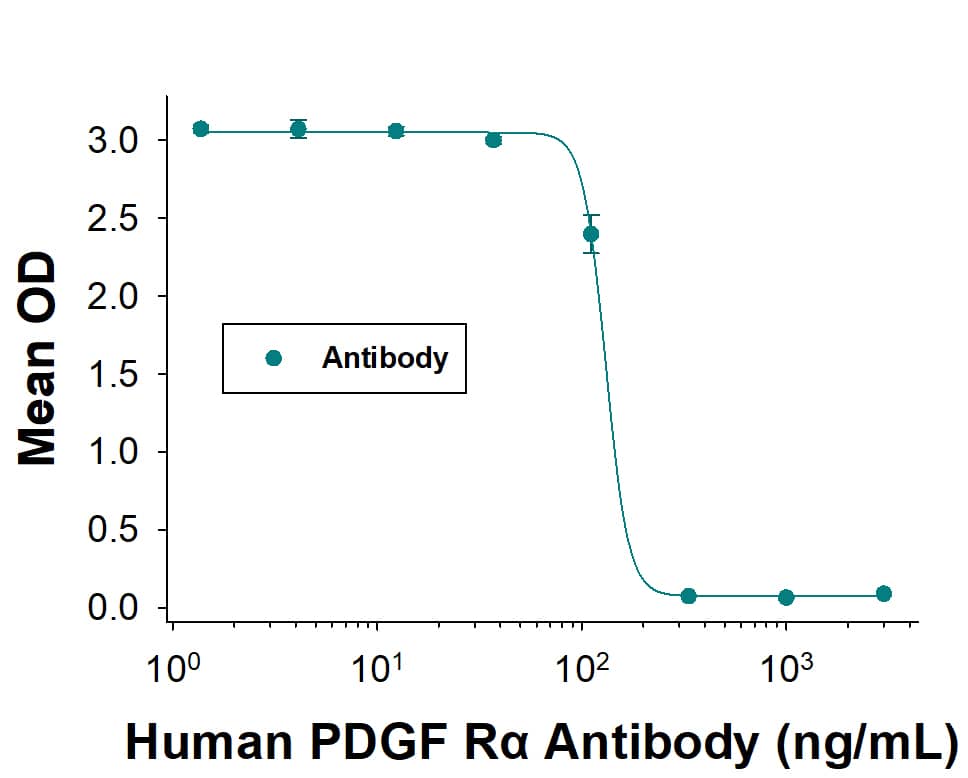
Neutralization by Human PDGF R alpha Antibody
In a functional ELISA, Human PDGF R alpha Antibody (Catalog # MAB322) blocks the binding of Recombinant Human PDGF R alpha Fc Chimera Protein (6765-PR) to Recombinant Human PDGF AA (221-AA). The Neutralization Dose (ND50) for this effect is typically 40.0-400 ng/mL.PDGF R alpha in Human Breast Cancer Tissue.
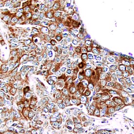
PDGF Ra was detected in immersion fixed paraffin-embedded sections of human breast cancer tissue using Mouse Anti-Human PDGF Ra Monoclonal Antibody (Catalog # MAB322) at 25 µg/mL overnight at 4 °C. Tissue was stained using the Anti-Mouse HRP-DAB Cell & Tissue Staining Kit (brown; Catalog # CTS002) and counterstained with hematoxylin (blue). Specific staining was localized to epithelial cells. View our protocol for Chromogenic IHC Staining of Paraffin-embedded Tissue Sections.Applications for Human PDGF R alpha Antibody
Immunohistochemistry
Sample: Immersion fixed paraffin-embedded sections of human breast cancer tissue
Western Blot
Sample: Recombinant Human PDGF R alpha (Catalog # 322-PR) under non-reducing conditions only
Neutralization
Reviewed Applications
Read 3 reviews rated 4.3 using MAB322 in the following applications:
Formulation, Preparation, and Storage
Purification
Reconstitution
Formulation
*Small pack size (-SP) is supplied either lyophilized or as a 0.2 µm filtered solution in PBS.
Shipping
Stability & Storage
- 12 months from date of receipt, -20 to -70 °C as supplied.
- 1 month, 2 to 8 °C under sterile conditions after reconstitution.
- 6 months, -20 to -70 °C under sterile conditions after reconstitution.
Background: PDGF R alpha
PDGF is a major serum mitogen that can exist as a homo- or heterodimeric protein consisting of disulfide-linked PDGF-A and PDGF-B chains. The PDGF-AA, PDGF‑BB and PDGF-AB isoforms have been shown to bind to two distinct cell surface PDGF receptors with different affinities. Whereas PDGF R alpha binds all three PDGF isoforms with high affinity, PDGF R beta binds PDGF‑BB and AB, but not PDGF-AA. Both PDGF R alpha and PDGF R beta are members of the class III subfamily of receptor tyrosine kinases (RTK) that also includes the receptors for M-CSF, SCF and Flt3 ligand. All class III RTKs are characterized by the presence of five immunoglobulin-like domains in their extracellular region and a split kinase domain in their intracellular region. PDGF binding induces receptor homo-and heterodimerization and signal transduction. The expression of the alpha and beta receptors is independently regulated in various cell types. Only PDGF R alpha is expressed in oligodendrocyte progenitor cells, mesothelial cell and liver endothelial cells. Soluble PDGF-R alpha has been detected in cell conditioned medium and human plasma. Recombinant soluble PDGF R alpha binds PDGF with high affinity and is a potent PDGF antagonist (1).
References
- Heldin, C.H. and L. Claesson-Welsh (1994) Guidebook to Cytokines and Their Receptors, Nicola, N.A. (ed) Oxford University Press, New York, NY p. 202.
Long Name
Alternate Names
Gene Symbol
Additional PDGF R alpha Products
Product Documents for Human PDGF R alpha Antibody
Product Specific Notices for Human PDGF R alpha Antibody
For research use only

