Human Phospho-ErbB2/Her2 (Y1248) Antibody
R&D Systems, part of Bio-Techne | Catalog # AF1768

Key Product Details
Validated by
Biological Validation
Species Reactivity
Validated:
Human
Cited:
Human
Applications
Validated:
Immunohistochemistry, Simple Western, Western Blot
Cited:
Westen Blot, Western Blot
Label
Unconjugated
Antibody Source
Polyclonal Rabbit IgG
Product Specifications
Immunogen
Phosphopeptide containing human ErbB2 Y1248 site
Specificity
Detects human ErbB2 when phosphorylated at Y1248.
Clonality
Polyclonal
Host
Rabbit
Isotype
IgG
Scientific Data Images for Human Phospho-ErbB2/Her2 (Y1248) Antibody
Detection of Human Phospho-ErbB2/Her2 (Y1248) by Western Blot.
Western blot shows lysates of MDA-MB-468 human breast cancer cell line and A431 human epithelial carcinoma cell line untreated (-) or treated (+) with 1 mM Pervanadate (PV) for 10 minutes or with 10 ng/mL Recombinant Human EGF (Catalog # 236-EG) for 5 minutes. PVDF membrane was probed with 0.25 µg/mL of Rabbit Anti-Human Phospho-ErbB2/Her2 (Y1248) Antigen Affinity-purified Polyclonal Antibody (Catalog # AF1768) followed by HRP-conjugated Anti-Rabbit IgG Secondary Antibody (Catalog # HAF008). A specific band was detected for Phospho-ErbB2/Her2 (Y1248) at approximately 170 kDa (as indicated). This experiment was conducted under reducing conditions and using Immunoblot Buffer Group 1.ErbB2/Her2 in Human Breast Cancer Tissue.
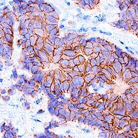
ErbB2/Her2 was detected in immersion fixed paraffin-embedded sections of human breast cancer tissue using Rabbit Anti-Human Phospho-ErbB2/Her2 (Y1248) Antigen Affinity-purified Polyclonal Antibody (Catalog # AF1768) at 0.3 µg/mL for 1 hour at room temperature followed by incubation with the Anti-Rabbit IgG VisUCyte™ HRP Polymer Antibody (VC003). Before incubation with the primary antibody, tissue was subjected to heat-induced epitope retrieval using Antigen Retrieval Reagent-Basic (VCTS021). Tissue was stained using DAB (brown) and counterstained with hematoxylin (blue). Specific staining was localized to plasma membrane. View our protocol for IHC Staining with VisUCyte HRP Polymer Detection Reagents.Detection of Human Phospho-ErbB2/Her2 (Y1248) by Simple WesternTM.
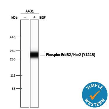
Simple Western lane view shows lysates of A431 human epithelial carcinoma cell line untreated (-) or treated (+) with 10 ng/mL Recombinant Human EGF (Catalog # 236-EG) for 5 minutes, loaded at 0.2 mg/mL. A specific band was detected for Phospho-ErbB2/Her2 (Y1248) at approximately 265 kDa (as indicated) using 5 µg/mL of Rabbit Anti-Human Phospho-ErbB2/Her2 (Y1248) Antigen Affinity-purified Polyclonal Antibody (Catalog # AF1768). This experiment was conducted under reducing conditions and using the 66-440 kDa separation system.Applications for Human Phospho-ErbB2/Her2 (Y1248) Antibody
Application
Recommended Usage
Immunohistochemistry
0.3-15 µg/mL
Sample: Immersion fixed paraffin-embedded sections of human breast cancer tissue
Sample: Immersion fixed paraffin-embedded sections of human breast cancer tissue
Simple Western
5 µg/mL
Sample: A431 human epithelial carcinoma cell line treated with Recombinant Human EGF (Catalog # 236-EG)
Sample: A431 human epithelial carcinoma cell line treated with Recombinant Human EGF (Catalog # 236-EG)
Western Blot
0.25 µg/mL
Sample: MDA‑MB‑468 human breast cancer cell line treated with Pervanadate (PV) and A431 human epithelial carcinoma cell line treated with Recombinant Human EGF (Catalog # 236-EG)
Sample: MDA‑MB‑468 human breast cancer cell line treated with Pervanadate (PV) and A431 human epithelial carcinoma cell line treated with Recombinant Human EGF (Catalog # 236-EG)
Formulation, Preparation, and Storage
Purification
Antigen Affinity-purified
Reconstitution
Reconstitute at 0.2 mg/mL in sterile PBS. For liquid material, refer to CoA for concentration.
Formulation
Lyophilized from a 0.2 μm filtered solution in PBS with Trehalose. *Small pack size (SP) is supplied either lyophilized or as a 0.2 µm filtered solution in PBS.
Shipping
Lyophilized product is shipped at ambient temperature. Liquid small pack size (-SP) is shipped with polar packs. Upon receipt, store immediately at the temperature recommended below.
Stability & Storage
Use a manual defrost freezer and avoid repeated freeze-thaw cycles.
- 12 months from date of receipt, -20 to -70 °C as supplied.
- 1 month, 2 to 8 °C under sterile conditions after reconstitution.
- 6 months, -20 to -70 °C under sterile conditions after reconstitution.
Background: ErbB2/Her2
References
- Coussens, L. et. al. (1985) Science 230:1132.
- Yamamoto, T. et. al. (1986) Nature 319:230.
- Kanai, Y. et. al. (1995) Biochem. Biophys. Res. Commun. 208:1067.
- Codony-Servat, J. et. al. (1999) Cancer Res. 59:1196.
- Carraway, K.L. 3rd et. al. (1994) J. Biol. Chem. 269:14303.
- Emkey, R. and C.R. Kahn (1997) J. Biol. Chem. 272:31172.
- Schaefer, G. et. al. (1999) J. Biol. Chem. 274:859.
- Schlessinger, J. (2000) Cell 103:211.
- Hellyer, N.J. et. al. (2001) J. Biol. Chem. 276:42153.
- Daly, R.J. (1999) Growth Factors 16:255.
Long Name
Receptor Tyrosine Protein Kinase ErbB2
Alternate Names
CD340, HER2, Neu Oncogene, NGL, TKR1
Gene Symbol
ERBB2
Additional ErbB2/Her2 Products
Product Documents for Human Phospho-ErbB2/Her2 (Y1248) Antibody
Product Specific Notices for Human Phospho-ErbB2/Her2 (Y1248) Antibody
For research use only
Loading...
Loading...
Loading...
Loading...
Loading...


