Human pIgR Antibody
R&D Systems, part of Bio-Techne | Catalog # AF2717


Key Product Details
Species Reactivity
Validated:
Cited:
Applications
Validated:
Cited:
Label
Antibody Source
Product Specifications
Immunogen
Lys19-Arg638
Accession # CAA51532
Specificity
Clonality
Host
Isotype
Endotoxin Level
Scientific Data Images for Human pIgR Antibody
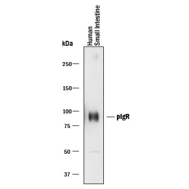
Detection of Human pIgR by Western Blot.
Western blot shows lysates of human small intestine tissue. PVDF membrane was probed with 0.2 µg/mL of Goat Anti-Human pIgR Antigen Affinity-purified Polyclonal Antibody (Catalog # AF2717) followed by HRP-conjugated Anti-Goat IgG Secondary Antibody (Catalog # HAF017). A specific band was detected for pIgR at approximately 85-100 kDa (as indicated). This experiment was conducted under reducing conditions and using Immunoblot Buffer Group 1.Applications for Human pIgR Antibody
Blockade of Receptor-ligand Interaction
Western Blot
Sample: Human small intestine tissue
Formulation, Preparation, and Storage
Purification
Reconstitution
Formulation
Shipping
Stability & Storage
- 12 months from date of receipt, -20 to -70 °C as supplied.
- 1 month, 2 to 8 °C under sterile conditions after reconstitution.
- 6 months, -20 to -70 °C under sterile conditions after reconstitution.
Background: pIgR
The human polymeric immunoglobulin receptor (pIgR; also known as membrane secretory component) is a 100 kDa type I transmembrane glycoprotein that is synthesized as a 764 amino acid (aa) precursor. It includes a signal sequence (aa 1-18), an extracellular region (aa 19-638), a transmembrane segment (aa 639-661), and a cytoplasmic domain (aa 662-764) (1-3). The extracellular region consists of five Ig-like domains and a sixth non-Ig domain that connects to the membrane region. pIgR is expressed on secretory epithelial cells of exocrine tissues. Immunoglobulin isotypes consist of two heavy (H) and two light (L) chains. For IgA and IgM, this H2L2 monomer can form larger polymers through association with a joining chain (J chain). The Fc regions of IgA and IgM have a carboxy-terminal extension called a secretory tailpiece that binds the J chain (4). pIgR functions as a carrier that transports IgA and IgM across epithelium (5). On the basolateral surface of epithelial cells, the receptor initially binds non-covalently to IgA via a docking site on the J chain. This initiates a rearrangement in which a disulfide bond forms between pIgR and an IgA heavy chain (2). The complexes are then internalized and transcytosed to the apical surface. A soluble covalent complex called secretory IgA (SIgA) is now generated by proteolytic cleavage of the sixth extracellular domain of pIgR and released into the lumen (6). This IgA-bound and proteolytically generated pIgR fragment is referred to as secretory component (SC). Notably, human pIgR transcytoses constitutively, with or without ligand, creating both bound and free, 78 kDa SC following cleavage (3). The extracellular region of pIgR is 64%, 65%, and 70% aa identical to the equivalent region in rat, mouse and porcine, respectively. The receptor component of the complex anchors the SIgA molecule to mucous (7). SIgA is a crucial component of the mucosal immune system serving to protect the large expanse of mucous membranes that form a barrier between the interior of the body and the external environment (8).
References
- Krajci, P. et al. (1989) Biochem. Biophys. Res. Commun. 158:783.
- Piskurich, J. et al. (1995) J. Immunol. 154:1735.
- Brandtzaeg, P. and F-E. Johansen (2001) Trends Immunol. 22:545.
- Braathen, R. et al. (2002) J. Biol. Chem. 277:42755.
- Ben-Hur, H. et al. (2004) Int. J. Mol. Med. 14:35.
- Asano, M. et al. (2004) Immunology 112:583.
- Phalipon, A. and B. Corthesy (2003) Trends Immunol. 24:55.
- Uren, T. et al. (2003) J. Immunol. 170:2531.
Long Name
Alternate Names
Gene Symbol
UniProt
Additional pIgR Products
Product Documents for Human pIgR Antibody
Product Specific Notices for Human pIgR Antibody
For research use only