Human PTPN14 Antibody
R&D Systems, part of Bio-Techne | Catalog # MAB4458

Key Product Details
Species Reactivity
Validated:
Cited:
Applications
Validated:
Cited:
Label
Antibody Source
Product Specifications
Immunogen
Leu329-Leu900
Accession # Q15678.2
Specificity
Clonality
Host
Isotype
Scientific Data Images for Human PTPN14 Antibody
Detection of Human PTPN14 by Western Blot.
Western blot shows lysates of DU145 human prostate carcinoma cell line, HT-29 human colon adenocarcinoma cell line, and HT-29 human colon adenocarcinoma cell line. PVDF membrane was probed with 1 µg/mL of Human PTPN14 Monoclonal Antibody (Catalog # MAB4458) followed by HRP-conjugated Anti-Mouse IgG Secondary Antibody (Catalog # HAF007). A specific band was detected for PTPN14 at approximately 135 kDa (as indicated). This experiment was conducted under reducing conditions and using Immunoblot Buffer Group 1.PTPN14 in Human Placenta.
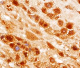
PTPN14 was detected in immersion fixed paraffin-embedded sections of human placenta using Human PTPN14 Monoclonal Antibody (Catalog # MAB4458) at 25 µg/mL overnight at 4 °C. Tissue was stained using the Anti-Mouse HRP-DAB Cell & Tissue Staining Kit (brown; Catalog # CTS002) and counterstained with hematoxylin (blue). View our protocol for Chromogenic IHC Staining of Paraffin-embedded Tissue Sections.Detection of Human PTPN14/PTPD2 by Western Blot
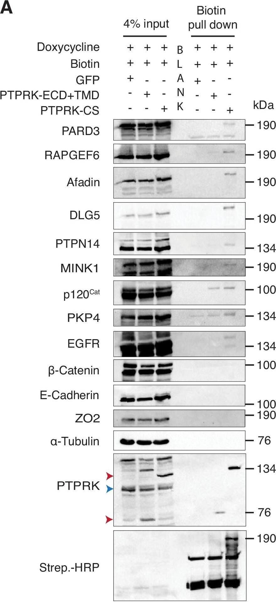
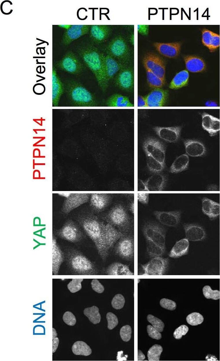
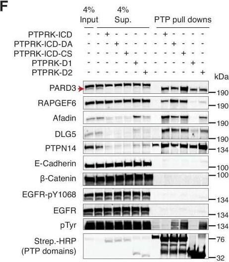
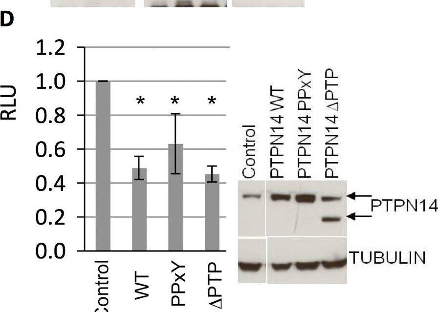
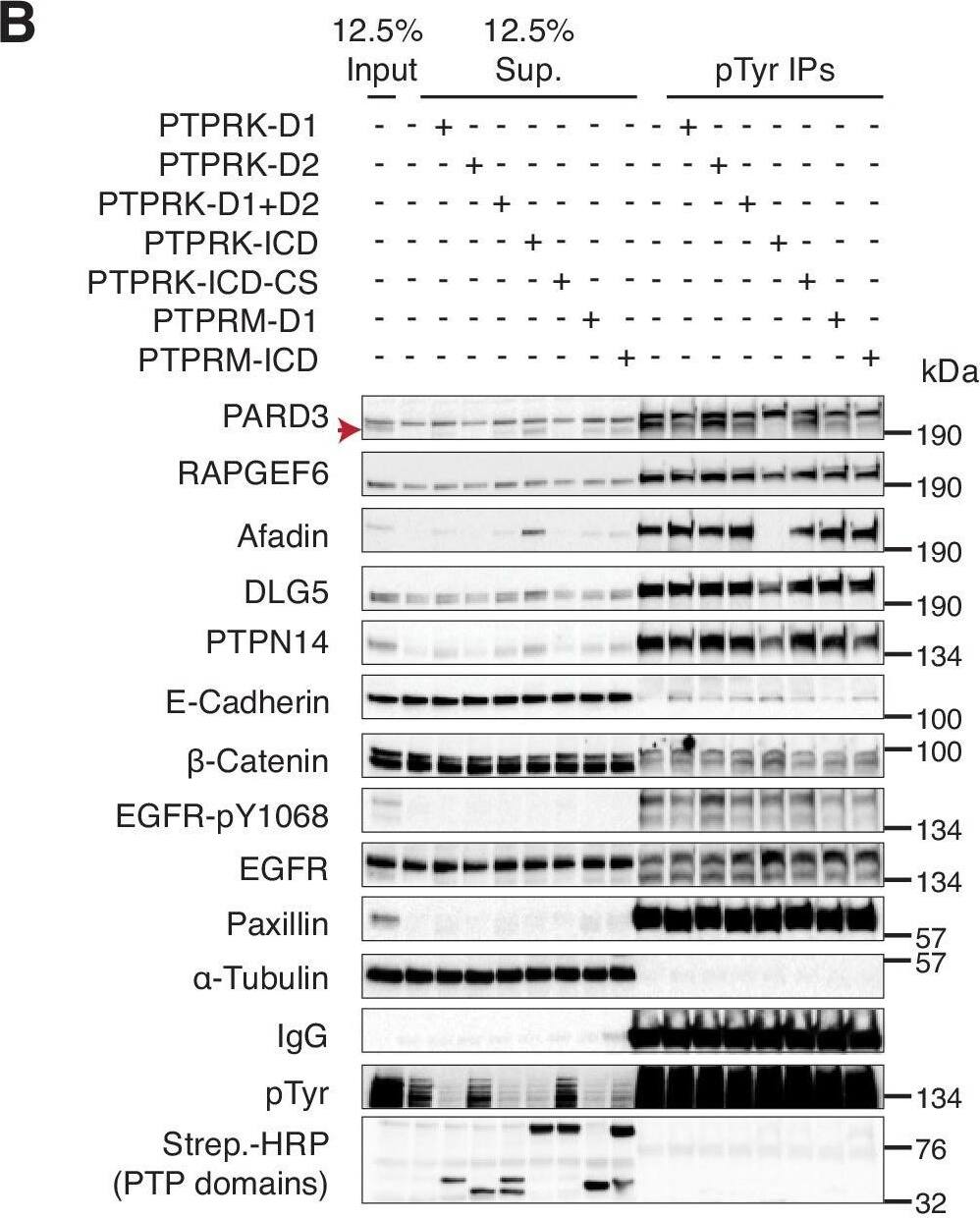
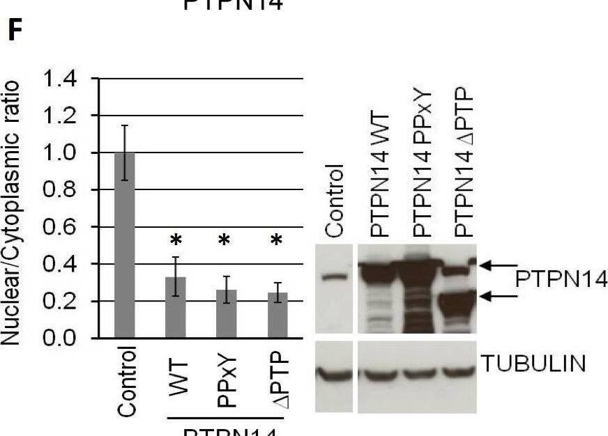
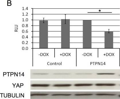
PTPRK interacts with candidate substrates in confluent MCF10A cells.(A) Representative immunoblot analysis of biotin pull downs from MCF10As expressing tGFP or PTPRK BioID constructs. See Materials and methods for details. Red and blue arrows indicate exogenous and endogenous PTPRK, respectively. (B) Quantification of BioID immunoblots. Green bars indicate the number of times a protein was enriched on PTPRK-C1089S.BirA*-Flag, compared to PTPRK.ECD +TMD.BirA*-Flag in separate experiments. Purple bars indicate the number of times a protein was not enriched or was not detected in any pull downs. n ≥ 1. (C) Schematic representation of PTPRK proximity-labeling by BioID. PTPRK extracellular domain homology model is based on PTPRM (PDB: 2V5Y; Aricescu et al., 2007). Proteins within the dotted lines were detected in pull downs from indicated BioID lysates. Proteins not detectably biotinylated are listed on the left. Proteins in bold and italics were previously identified as PTPRK interactors using BioID in HEK293 cells (St-Denis et al., 2016). See also Figure 4—figure supplement 1.Localization of PTPRK BioID proteins.(A) MCF10As with stably integrated doxycycline-inducible expression constructs (PTPRK-ECD +TMD-BirA*-Flag and PTPRK-C1089s-BirA*-Flag) were treated with 150 ng/ml and 500 ng/ml doxycycline, respectively, and immunostained using an anti-Flag antibody. F-actin and nuclei were stained with phalloidin and Hoechst, respectively. Scale bars = 50 µm. (B) Representative immunoblot analysis of biotin pull downs from MCF10As expressing tGFP or PTPRK BioID constructs. See Materials and methods for details. Red and blue arrows indicate exogenous and endogenous PTPRK, respectively. * residual ABLIM3 signal. Image collected and cropped by CiteAb from the following open publication (https://pubmed.ncbi.nlm.nih.gov/30924770), licensed under a CC-BY license. Not internally tested by R&D Systems.Applications for Human PTPN14 Antibody
Immunohistochemistry
Sample: Immersion fixed paraffin-embedded human placenta
Western Blot
Sample: DU145 human prostate carcinoma cell line, HT-29 human colon adenocarcinoma cell line, and HT-29 human colon adenocarcinoma cell line
Reviewed Applications
Read 3 reviews rated 4 using MAB4458 in the following applications:
Formulation, Preparation, and Storage
Purification
Reconstitution
Formulation
Shipping
Stability & Storage
- 12 months from date of receipt, -20 to -70 °C as supplied.
- 1 month, 2 to 8 °C under sterile conditions after reconstitution.
- 6 months, -20 to -70 °C under sterile conditions after reconstitution.
Background: PTPN14
Protein Tyrosine Phosphatase, Non-receptor type 14 (PTPN14), also called Phosphatase with Ezrin Domain (PEZ) and PTP36, is a 135 kDa cytosolic protein that dephosphorylates tyrosine residues in proteins. PTPN14’s Ezrin domain is homologous to erythrocyte Band 4.1 protein, targeting the phosphatase to cytoskeletal elements such as actin. PTPN14 associates with beta-catenin in adhesion zones in cells that are touching, but is nuclear in isolated, proliferating cells. Loss-of-function mutations in PTPN14 are found in 26% of colorectal cancers, suggesting it is a tumor suppressor.
Long Name
Alternate Names
Gene Symbol
UniProt
Additional PTPN14 Products
Product Documents for Human PTPN14 Antibody
Product Specific Notices for Human PTPN14 Antibody
For research use only
