Human Rab27a Antibody
R&D Systems, part of Bio-Techne | Catalog # MAB7245

Key Product Details
Validated by
Species Reactivity
Validated:
Cited:
Applications
Validated:
Cited:
Label
Antibody Source
Product Specifications
Immunogen
Ser135-Ala218
Accession # P51159
Specificity
Clonality
Host
Isotype
Scientific Data Images for Human Rab27a Antibody
Detection of Human Rab27a by Western Blot.
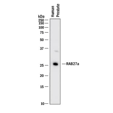
Western blot shows lysates of human prostate tissue. PVDF membrane was probed with 2 µg/mL of Rabbit Anti-Human Rab27a Monoclonal Antibody (Catalog # MAB7245) followed by HRP-conjugated Anti-Rabbit IgG Secondary Antibody (HAF008). A specific band was detected for Rab27a at approximately 26 kDa (as indicated). This experiment was conducted under reducing conditions and using Immunoblot Buffer Group 1.Detection of Human Rab27a by Simple WesternTM.
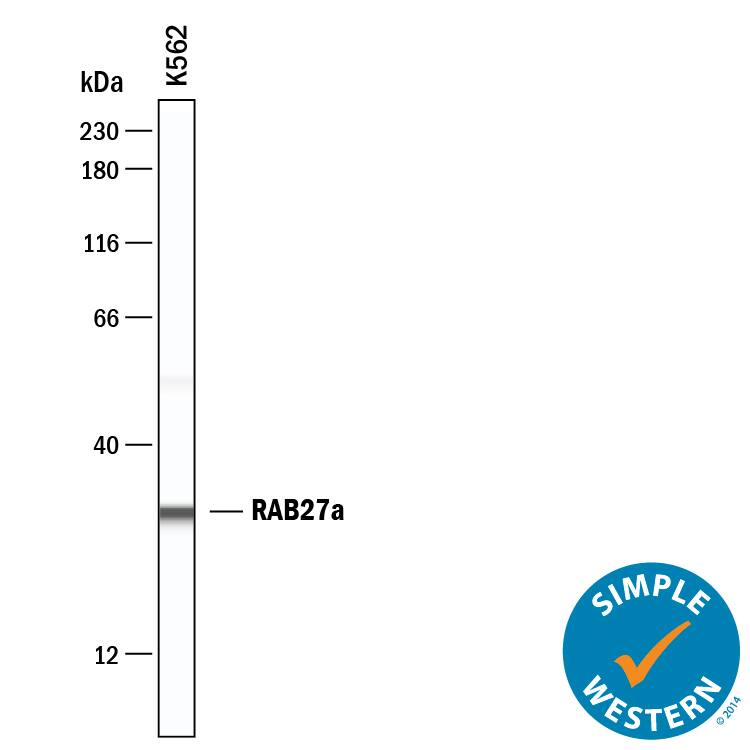
Simple Western shows lysates of Exosome Standards (K562) (NBP2-49864) and SK-Mel-28 human malignant melanoma cell line, loaded at 0.5 mg/ml. A specific band was detected for Rab27a at approximately 32 kDa (as indicated) using 20 µg/mL of Rabbit Anti-Human Rab27a Monoclonal Antibody (Catalog # MAB7245). This experiment was conducted under reducing conditions and using the 12-230 kDa separation system.Rab27a in SK‑Mel‑28 Human Cell Line.
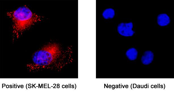
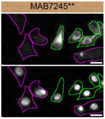
Rab27a was detected in immersion fixed SK-Mel-28 human malignant melanoma cell line (left panel; positive staining) and Daudi human Burkitt's lymphoma cell line (right panel; negative staining) using Rabbit Anti-Human Rab27a Monoclonal Antibody (Catalog # MAB7245) at 3 µg/mL for 3 hours at room temperature. Cells were stained using the NorthernLights™ 557-conjugated Anti-Rabbit IgG Secondary Antibody (red; NL004) and counterstained with DAPI (blue). Specific staining was localized to cytoplasm. View our protocol for Fluorescent ICC Staining of Cells on Coverslips.Applications for Human Rab27a Antibody
Immunocytochemistry
Sample: Immersion fixed SK-Mel-28 human malignant melanoma cell line
Simple Western
Sample: Exosome Standards (K562) (Catalog # NBP2-49864), SK-Mel-28 human malignant melanoma cell line and K562 human chronic myelogenous leukemia cell line
Western Blot
Sample: Human prostate tissue
Reviewed Applications
Read 1 review rated 5 using MAB7245 in the following applications:
Formulation, Preparation, and Storage
Purification
Reconstitution
Formulation
*Small pack size (-SP) is supplied either lyophilized or as a 0.2 µm filtered solution in PBS.
Shipping
Stability & Storage
- 12 months from date of receipt, -20 to -70 °C as supplied.
- 1 month, 2 to 8 °C under sterile conditions after reconstitution.
- 6 months, -20 to -70 °C under sterile conditions after reconstitution.
Background: Rab27a
RAB27A (Ras-related protein Rab 27A; also GTP-binding protein Ram) is a 27-28 kDa member of the Rab27 subfamily, Rab family, Small GTPase superfamily of proteins. It is widely expressed, and found in cells diverse as mast cells, cytotoxic T cells, melanocytes, retinal pigment epithelium and pancreatic beta-cells. RAB27A plays a key role in the secretion of specialized lysosomes termed secretory lysosomes. In melanocytes, for example, RAB27A is incorporated into the melanosome membrane where it serves as a docking factor for melanophilin and myosin-Va, regulating melanosome transport to, and concentration at, sites of release. Human RAB27A is 221 amino acids (aa) in length. It contains multiple Rab family and subfamily motifs, and concludes with a C-terminal CXC prenylation sequence (aa 219‑221). There is one potential splice variant that shows a deletion of aa 146-153. Over aa 135-218, human RAB27A shares 92% and 94% aa sequence identity with mouse Rab27A and rat RAB27A, respectively.
Long Name
Alternate Names
Entrez Gene IDs
Gene Symbol
UniProt
Additional Rab27a Products
Product Documents for Human Rab27a Antibody
Product Specific Notices for Human Rab27a Antibody
For research use only

