Human Resistin Antibody
R&D Systems, part of Bio-Techne | Catalog # AF1359


Key Product Details
Species Reactivity
Validated:
Human
Cited:
Human
Applications
Validated:
Western Blot
Cited:
ELISA Development, Immunocytochemistry, Mass Spectometry, Western Blot
Label
Unconjugated
Antibody Source
Polyclonal Goat IgG
Product Specifications
Immunogen
E. coli-derived recombinant human Resistin
Ser17-Pro108
Accession # Q9HD89
Ser17-Pro108
Accession # Q9HD89
Specificity
Detects human Resistin in direct ELISAs and Western blots. In these formats, this antibody shows less than 2% cross-reactivity with rmResistin, rmRELM alpha and rmRELM beta.
Clonality
Polyclonal
Host
Goat
Isotype
IgG
Scientific Data Images for Human Resistin Antibody
Detection of Human Resistin by Block/Neutralize
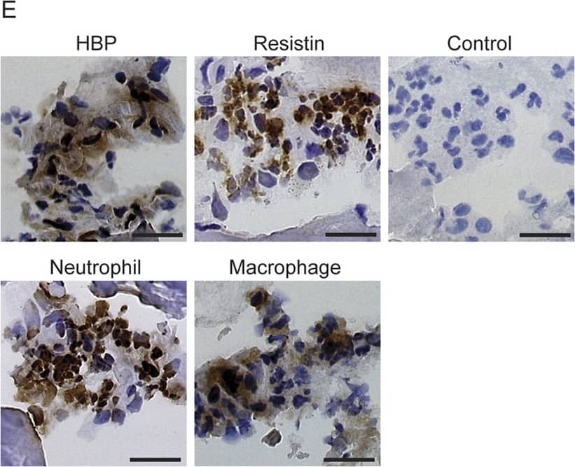
Systemic and local HBP and resistin responses in septic patients.HBP and resistin levels in plasma collected from patients on the day of inclusion were measured with ELISA, for details see Experimental procedures. (A,B) HBP and resistin levels in plasma from patients with severe sepsis/septic shock (n = 88) or non-infected critically ill patients (n = 31). (C) Plasma HBP and resistin levels in severe sepsis/septic shock patients with confirmed Gram-positive (G+) or Gram-negative (G−) infections (n = 48). (D) Systemic HBP and resistin in STSS patients (n = 8). The correlation was determined by Pearson’s correlation test, indicated by p and r-values. (E) HBP, resistin, macrophages and neutrophils were analysed by immunohistochemical staining of cryosections of snap-frozen tissue biopsies (n = 9) from patients with severe soft tissue infections caused by GAS. A control staining where the primary antibody was omitted was also performed (control). A representative tissue biopsy is shown. The scale bar indicates 50 μm. (F) The stainings were evaluated by microscope and acquired computerized image analysis (ACIA), for details see Experimental procedures. Significant correlation was determined by Spearman test, indicated by p- and r-values. Image collected and cropped by CiteAb from the following open publication (https://www.nature.com/articles/srep21288), licensed under a CC-BY license. Not internally tested by R&D Systems.Detection of Human Resistin by Block/Neutralize
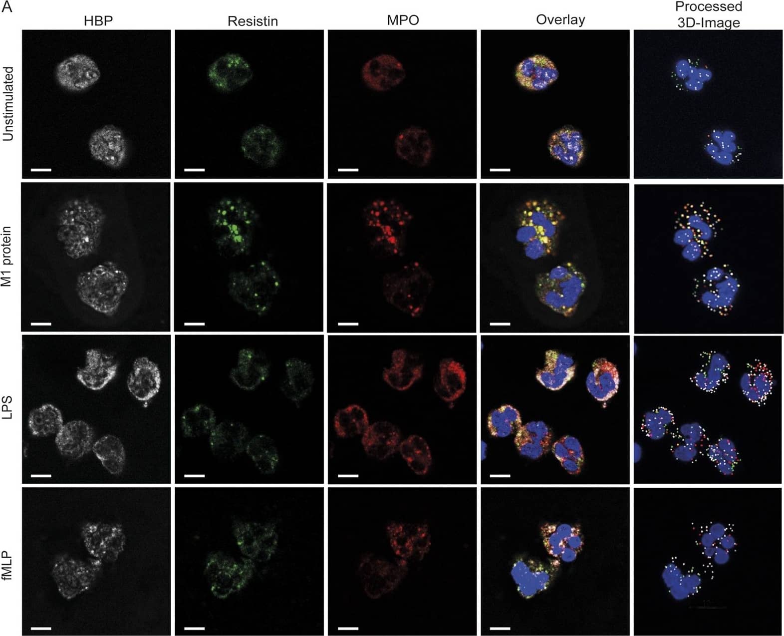
Subcellular localization of HBP and resistin in neutrophils.Neutrophils from healthy donors were isolated and stimulated with LPS (50 ng/ml), fMLP (5 μg/ml) or streptococcal M1-protein (1 μg/ml) for 2 h. The neutrophils were immunoflourescently stained for HBP (white), resistin (green) and MPO (red), the cell nuclei were visualized using DAPI (blue). Using scanning laser confocal microscopy, Z-stacks of the cells were acquired and vesicles positive for HBP, resistin or MPO identified using the Imaris image analysis software (processed 3D-Image). The degree of colocalization between the above-mentioned markers in the processed images was then quantified using Imaris analysis software. (A) Shows images of cells from a high responder. Bars indicate 5 μm. (B–D) Percentage of co-localization between factors as indicated. (E) Differences in co-localization in neutrophils unstimulated or stimulated with either M1-protein or LPS. Statistical significant differences were determined using the Mann Whitney test, **P < 0.01. Data represent results obtained from multiple field analyses of two experiments using different donors. Image collected and cropped by CiteAb from the following open publication (https://www.nature.com/articles/srep21288), licensed under a CC-BY license. Not internally tested by R&D Systems.Applications for Human Resistin Antibody
Application
Recommended Usage
Western Blot
0.1 µg/mL
Sample: Recombinant Human Resistin
Sample: Recombinant Human Resistin
Reviewed Applications
Read 2 reviews rated 4.5 using AF1359 in the following applications:
Formulation, Preparation, and Storage
Purification
Antigen Affinity-purified
Reconstitution
Reconstitute at 0.2 mg/mL in sterile PBS. For liquid material, refer to CoA for concentration.
Formulation
Lyophilized from a 0.2 μm filtered solution in PBS with Trehalose. *Small pack size (SP) is supplied either lyophilized or as a 0.2 µm filtered solution in PBS.
Shipping
Lyophilized product is shipped at ambient temperature. Liquid small pack size (-SP) is shipped with polar packs. Upon receipt, store immediately at the temperature recommended below.
Stability & Storage
Use a manual defrost freezer and avoid repeated freeze-thaw cycles.
- 12 months from date of receipt, -20 to -70 °C as supplied.
- 1 month, 2 to 8 °C under sterile conditions after reconstitution.
- 6 months, -20 to -70 °C under sterile conditions after reconstitution.
Background: Resistin
Alternate Names
ADSF, FIZZ3, HXCP1, RETN
Gene Symbol
RETN
UniProt
Additional Resistin Products
Product Documents for Human Resistin Antibody
Product Specific Notices for Human Resistin Antibody
For research use only
Loading...
Loading...
Loading...
Loading...
