Human Semaphorin 3A Antibody
R&D Systems, part of Bio-Techne | Catalog # MAB1250


Key Product Details
Species Reactivity
Validated:
Cited:
Applications
Validated:
Cited:
Label
Antibody Source
Product Specifications
Immunogen
Lys26-Val771 (Arg555Ala, Arg552Ala)
Accession # Q14563
Specificity
Clonality
Host
Isotype
Scientific Data Images for Human Semaphorin 3A Antibody
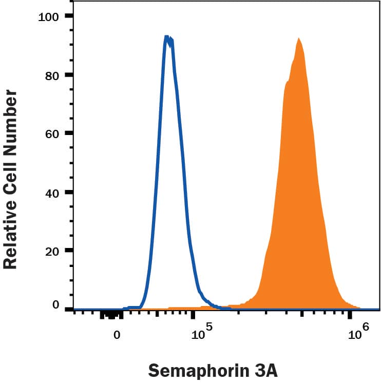
Detection of Semaphorin 3A in U118-MG cells by Flow Cytometry
U118-MG cells were stained with Mouse Anti-Human Semaphorin 3A Monoclonal Antibody (Catalog # MAB1250, filled histogram) or isotype control antibody (Catalog # MAB0041, open histogram) followed by Fluorescein-conjugated Anti-Mouse IgG Secondary Antibody (Catalog # F0103B). To facilitate intracellular staining, cells were fixed with Flow Cytometry Fixation Buffer (Catalog # FC004) and permeabilized with Flow Cytometry Permeabilization/Wash Buffer I (Catalog # FC005). View our protocol for Staining Intracellular Molecules.Applications for Human Semaphorin 3A Antibody
CyTOF-ready
Intracellular Staining by Flow Cytometry
Sample: U118-MG cells
Western Blot
Sample: Recombinant Human Semaphorin 3A Fc Chimera (Catalog # 1250-S3)
Reviewed Applications
Read 1 review rated 5 using MAB1250 in the following applications:
Formulation, Preparation, and Storage
Purification
Reconstitution
Formulation
*Small pack size (-SP) is supplied either lyophilized or as a 0.2 µm filtered solution in PBS.
Shipping
Stability & Storage
- 12 months from date of receipt, -20 to -70 °C as supplied.
- 1 month, 2 to 8 °C under sterile conditions after reconstitution.
- 6 months, -20 to -70 °C under sterile conditions after reconstitution.
Background: Semaphorin 3A
The Semaphorins constitute a large family of secreted, GPI-anchored and transmembrane cell signaling molecules. Depending on their domain organization and species origin, these proteins can be classified into eight groups. To date, at least 19 vertebrate Semaphorins belonging to five groups (class 3 through 7) have been identified. All Semaphorins contain a conserved, 500 amino acid (aa) Sema domain at the amino terminus. Semaphorins are best known for their roles in axon guidance during neuronal development. Semaphorins are also expressed in non-neuronal tissues and are involved in angiogenesis, hematopoiesis, organogenesis, and the regulation of immune functions (1, 2).
Class 3 Semaphorins (Sema3) are secreted proteins containing a Sema domain, an immunoglobulin c2-like domain and a basic domain near the carboxyl tail. Sema3A (also referred to as SemaIII, SemD and Collapsin) cDNA predicts a 771 aa precursor protein with a putative 25 aa signal peptide (1‑3). Bioactive Sema3A is a disulfide-linked dimer (4). The bioactivity is increased after proteolytic processing by a furin-like endoprotease near the carboxy-terminus (1). The functional receptor complex for Sema3 is composed of two distinct transmembrane proteins: Neuropilin-1 (Npn-1) and Plexin-A. Npn-1 binds directly to Sema3A with high-affinity and confers specificity. Plexin-A interacts with Npn-1 to increase the affinity of the complex for Sema3A and serves as the signaling subunit in the receptor complex (1, 2, 5).
References
- Nakamura, F. et al. (2000) J Neurobiol. 44:219.
- Goshima, Y. et al. (2002) J. Clin. Invest. 109:993.
- Kolodkin, A.L. et al. (1993) Cell 75:1389.
- Koppel, A.M. et al. (1998) J. Biol. Chem. 273:15708.
- Yu, T.W. et al. (2001) Nature Neurosci. Supplement 4:1169.
- Luo Y. et al. (1993) Cell 75:217.
Alternate Names
Entrez Gene IDs
Gene Symbol
UniProt
Additional Semaphorin 3A Products
Product Documents for Human Semaphorin 3A Antibody
Product Specific Notices for Human Semaphorin 3A Antibody
For research use only