Human SHB Antibody
R&D Systems, part of Bio-Techne | Catalog # AF7036

Key Product Details
Species Reactivity
Applications
Label
Antibody Source
Product Specifications
Immunogen
Ala2-Phe128
Accession # Q15464
Specificity
Clonality
Host
Isotype
Scientific Data Images for Human SHB Antibody
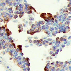
SHB in Human Ovarian Cancer Tissue.
SHB was detected in immersion fixed paraffin-embedded sections of human ovarian cancer tissue using Goat Anti-Human SHB Antigen Affinity-purified Polyclonal Antibody (Catalog # AF7036) at 10 µg/mL overnight at 4 °C. Tissue was stained using the Anti-Goat HRP-DAB Cell & Tissue Staining Kit (brown; Catalog # CTS008) and counterstained with hematoxylin (blue). Specific staining was localized to cytoplasm in epithelial cells. View our protocol for Chromogenic IHC Staining of Paraffin-embedded Tissue Sections.Applications for Human SHB Antibody
Immunohistochemistry
Sample: Immersion fixed paraffin-embedded sections of human ovarian cancer tissue
Formulation, Preparation, and Storage
Purification
Reconstitution
Formulation
Shipping
Stability & Storage
- 12 months from date of receipt, -20 to -70 °C as supplied.
- 1 month, 2 to 8 °C under sterile conditions after reconstitution.
- 6 months, -20 to -70 °C under sterile conditions after reconstitution.
Background: SHB
SHB (SH2 homology protein in B/ beta-cells) is a 55-59 kDa cytoplasmic adaptor protein that serves as a link between phosphotyrosine residues and downstream signaling pathways. SHB is ubiquitously expressed, and binds tyrosine kinase receptors such as FGFR1, VEGFR2, and the TCR ( zeta-chain) following their activation. It is highly modular, and through a variety of motifs, is able to bind multiple, structurally unrelated proteins that collectively generate a signal transduction network. Human SHB is 509 amino acids (aa) in length. It contains an N-terminal Pro-rich region that binds SH3 domain-containing proteins (aa 19-45), a central PTB domain that binds select aa motifs, and a C-terminal SH2 domain that interacts with phosphotyrosines (aa 410-504). There are at least nine utilized phosphorylation sites. There are two isoform variants. One is 66-68 kDa in size, and possesses an 87 aa Pro-rich N-terminal extension. This MW may increase to 77-80 kDa following posttranslational processing. A second variant shows an 18 aa substitution for aa 280-509. Over aa 2-128, human SHB shares 91% aa identity with mouse SHB.
Long Name
Alternate Names
Gene Symbol
UniProt
Additional SHB Products
Product Documents for Human SHB Antibody
Product Specific Notices for Human SHB Antibody
For research use only
