Human SOD3/EC-SOD Antibody
R&D Systems, part of Bio-Techne | Catalog # AF3420

Key Product Details
Species Reactivity
Validated:
Cited:
Applications
Validated:
Cited:
Label
Antibody Source
Product Specifications
Immunogen
Trp19-Ala240
Accession # P08294
Specificity
Clonality
Host
Isotype
Scientific Data Images for Human SOD3/EC-SOD Antibody
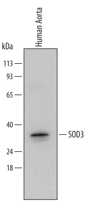
Detection of Human SOD3/EC‑SOD by Western Blot.
Western blot shows lysates of human aorta tissue. PVDF Membrane was probed with 0.2 µg/mL of Goat Anti-Human SOD3/EC-SOD Antigen Affinity-purified Polyclonal Antibody (Catalog # AF3420) followed by HRP-conjugated Anti-Goat IgG Secondary Antibody (Catalog # HAF017). A specific band was detected for SOD3/EC-SOD at approximately 30 kDa (as indicated). This experiment was conducted under reducing conditions and using Immunoblot Buffer Group 2.SOD3/EC‑SOD in Human Kidney.
SOD3/EC-SOD was detected in immersion fixed paraffin-embedded sections of human kidney using Goat Anti-Human SOD3/EC-SOD Antigen Affinity-purified Polyclonal Antibody (Catalog # AF3420) at 5 µg/mL overnight at 4 °C. Tissue was stained using the Anti-Goat HRP-DAB Cell & Tissue Staining Kit (brown; Catalog # CTS008) and counterstained with hematoxylin (blue). Specific staining was localized to the cytoplasm of epithelial cells in convoluted tubules. View our protocol for Chromogenic IHC Staining of Paraffin-embedded Tissue Sections.Detection of Human SOD3/EC-SOD by Simple WesternTM.
Simple Western lane view shows lysates of human kidney tissue, loaded at 0.2 mg/mL. A specific band was detected for SOD3/EC-SOD at approximately 38 kDa (as indicated) using 10 µg/mL of Goat Anti-Human SOD3/EC-SOD Antigen Affinity-purified Polyclonal Antibody (Catalog # AF3420) followed by 1:50 dilution of HRP-conjugated Anti-Goat IgG Secondary Antibody (Catalog # HAF109). This experiment was conducted under reducing conditions and using the 12-230 kDa separation system.Applications for Human SOD3/EC-SOD Antibody
Immunohistochemistry
Sample: Immersion fixed paraffin-embedded sections of human kidney
Simple Western
Sample: Human kidney tissue
Western Blot
Sample: Human aorta tissue
Reviewed Applications
Read 1 review rated 5 using AF3420 in the following applications:
Formulation, Preparation, and Storage
Purification
Reconstitution
Formulation
Shipping
Stability & Storage
- 12 months from date of receipt, -20 to -70 °C as supplied.
- 1 month, 2 to 8 °C under sterile conditions after reconstitution.
- 6 months, -20 to -70 °C under sterile conditions after reconstitution.
Background: SOD3/EC-SOD
Superoxide Dismutases (SODs), originally identified as Indophenoloxidase (IPO), are enzymes that catalyze the conversion of naturally-occuring, but harmful, superoxide radicals into molecular oxygen and hydrogen peroxide. Superoxide Dismutases 3, SOD3, also known as extracellular (EC) SOD, is tetrameric glycoprotein with an apparent subunit molecular weight of about 30 kDa. Three isoenzymes of SOD have been identified and are functionally related but have very modest sequence homology. SOD3 shares 23% and 17% sequence identity with SOD1 and SOD2, respectively. SOD3 shares ~64% sequence homology with mouse and rat SOD3. Like SOD1, SOD3 binds one Cu2+ and Zn2+ ions per subunit but differs in sequence and tissue distribution. SOD3 is a secretory protein and is synthesized with a putative 18-amino acid signal peptide preceding the 222 amino acids in the mature SOD3. SOD3 is found in plasma, lymph, and synovial fluid as well as in tissues. SOD3 binds on the surface of endothelial cells through the heparan sulfate proteoglycan and eliminates the oxygen radicals from the NADP-dependent oxidative system of neutrophils.
Long Name
Alternate Names
Gene Symbol
UniProt
Additional SOD3/EC-SOD Products
Product Documents for Human SOD3/EC-SOD Antibody
Product Specific Notices for Human SOD3/EC-SOD Antibody
For research use only


