Human SOX3 Antibody
R&D Systems, part of Bio-Techne | Catalog # AF2569

Key Product Details
Species Reactivity
Validated:
Human
Cited:
Mouse
Applications
Validated:
Immunocytochemistry, Western Blot
Cited:
Flow Cytometry, Immunocytochemistry, Immunohistochemistry, Immunohistochemistry-Frozen, Immunoprecipitation
Label
Unconjugated
Antibody Source
Polyclonal Goat IgG
Product Specifications
Immunogen
E. coli-derived recombinant human SOX3
Met1-Leu230
Accession # P41225
Met1-Leu230
Accession # P41225
Specificity
Detects human SOX3 in direct ELISAs and Western blots. In direct ELISAs and Western blots, less than 1% cross-reactivity with recombinant human (rh) SOX2, rhSOX7, rhSOX10, and rhSOX17 is observed.
Clonality
Polyclonal
Host
Goat
Isotype
IgG
Scientific Data Images for Human SOX3 Antibody
SOX3 in NTera‑2 Human Cell Line.
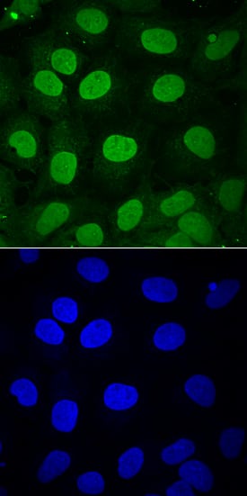
SOX3 was detected in immersion fixed NTera-2 human testicular embryonic carcinoma cell line differentiation with retinoic acid using Goat Anti-Human SOX3 Antigen Affinity-purified Polyclonal Antibody (Catalog # AF2569) at 10 µg/mL for 3 hours at room temperature. Cells were stained using the NorthernLights™ 493-conjugated Anti-Goat IgG Secondary Antibody (green, upper panel; NL003) and counterstained with DAPI (blue, lower panel). View our protocol for Fluorescent ICC Staining of Cells on Coverslips.Detection of SOX3 in U-251 MG cells (positive) and HDLM-2 cells (negative).
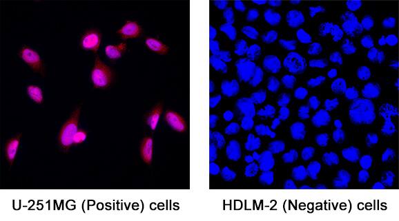
SOX3 was detected in immersion fixed U-251 MG human glioblastoma cells (positive) and absent in HDLM-2 human Hodgkin’s lymphoma cells (negative) using Goat Anti-Human SOX3 Antigen Affinity-purified Polyclonal Antibody (Catalog # AF2569) at 15 µg/mL for 3 hours at room temperature. Cells were stained using the NorthernLights™ 557-conjugated Anti-Goat IgG Secondary Antibody (red; Catalog # NL001) and counterstained with DAPI (blue). Specific staining was localized to cell nuclei. View our protocol for Fluorescent ICC Staining of Cells on Coverslips.Detection of SOX3 by Western Blot
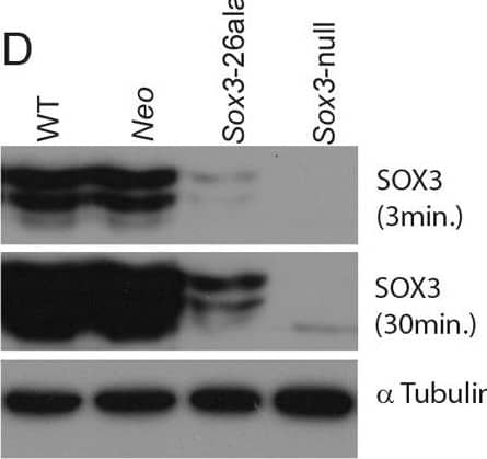
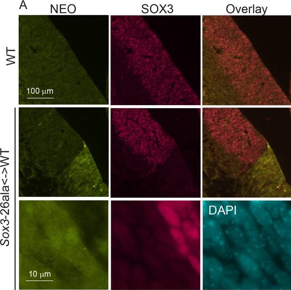
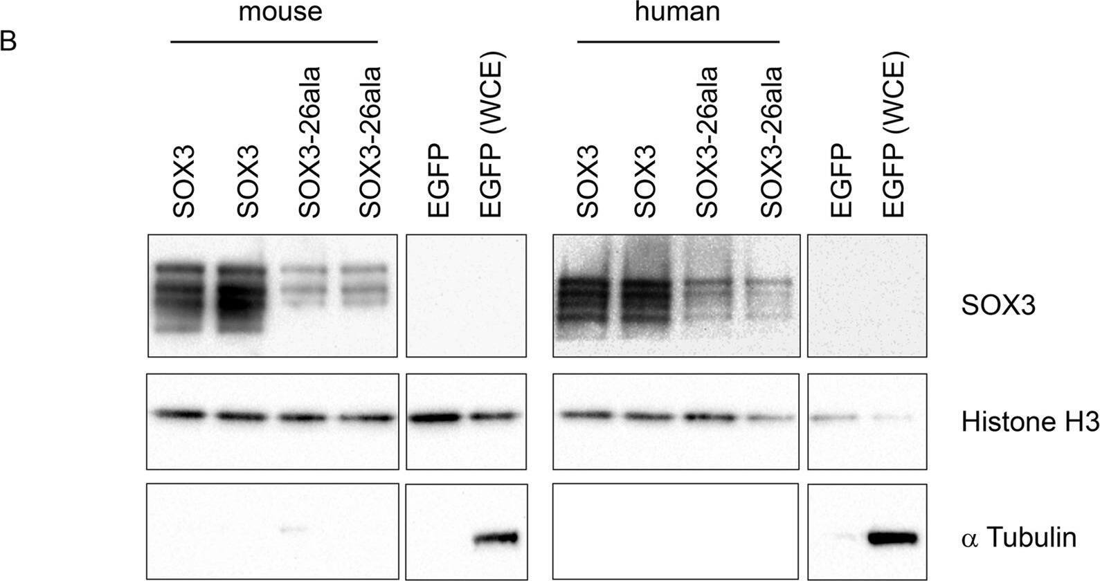
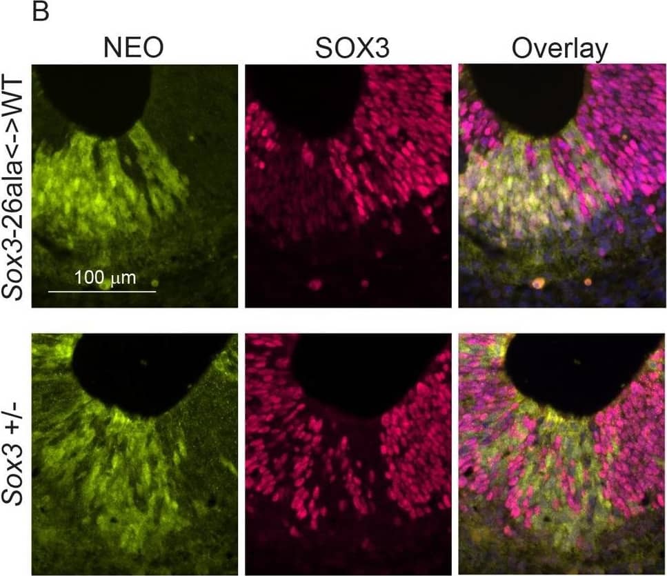
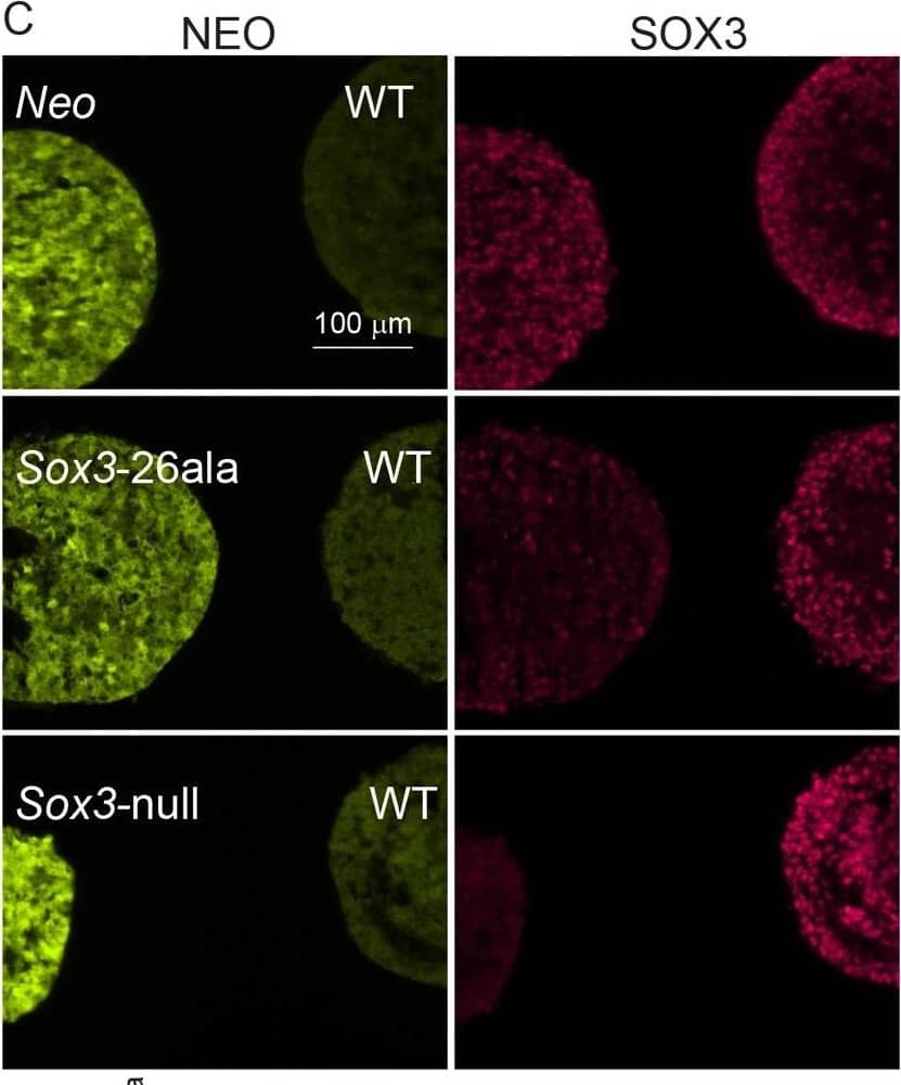
Transcription is unaffected but protein is cleared from mutant cells.A) SOX3 protein is present in every WT cell (NEO−) of the 13.5 dpc telencephalic ventricular zone but virtually absent from equivalent tissue derived from Sox3-26ala cells (NEO+). B) Comparison of SOX3 immunostaining on Sox3-null cells (from a 14.5 dpc +/− embryo) and Sox3-26ala expressing cells (from a Sox3-26ala <-> WT chimera) confirming that the antibody is SOX3-specific and that the Sox3-26ala expressing cells exhibit a low level of residual nuclear protein. C) WT, Neo, Sox3-26ala and Sox3-null ES cells were differentiated for 5 days in CDM as multi-cellular bodies. Rare SOX3 positive cells were detected in Sox3-26ala CDMs while the majority of cells had low SOX3 protein levels in comparison to neighbouring WT CDM bodies processed on the same slide. D–E) WT, Neo, Sox3-26ala and Sox3-null ES cells were grown in N2B27 for 4 days to form neural progenitors. Western blotting for SOX3 reveals a dramatic reduction of protein in Sox3-26ala cells (D); 3 and 30 minute exposures are shown. E) Transcript levels of Sox3 are not affected in Sox3-26ala cells as determined by qPCR. Three experimental replicates are shown. Data was normalised to Sox3 levels inSox3-Neo control cells and error bars represent SEM. F) ISH confirms that Sox3 transcript is present at comparable levels in ventricular zone cells at 13.5 dpc derived from both WT (Neo−) and Sox3-26ala (Neo+) cells. ISH performed on adjacent 10 µm coronal sections of 13.5 dpc chimeric telencephalon. Image collected and cropped by CiteAb from the following open publication (https://pubmed.ncbi.nlm.nih.gov/23505376), licensed under a CC-BY license. Not internally tested by R&D Systems.Applications for Human SOX3 Antibody
Application
Recommended Usage
Immunocytochemistry
5-15 µg/mL
Sample: Immersion fixed NTera-2 human testicular embryonic carcinoma cell line, U-251 MG human glioblastoma cells (positive) and HDLM-2 human Hodgkin's lymphoma cells (negative)
Sample: Immersion fixed NTera-2 human testicular embryonic carcinoma cell line, U-251 MG human glioblastoma cells (positive) and HDLM-2 human Hodgkin's lymphoma cells (negative)
Western Blot
0.1 µg/mL
Sample: Recombinant Human SOX3
Sample: Recombinant Human SOX3
Formulation, Preparation, and Storage
Purification
Antigen Affinity-purified
Reconstitution
Reconstitute at 0.2 mg/mL in sterile PBS. For liquid material, refer to CoA for concentration.
Formulation
Lyophilized from a 0.2 μm filtered solution in PBS with Trehalose. *Small pack size (SP) is supplied either lyophilized or as a 0.2 µm filtered solution in PBS.
Shipping
Lyophilized product is shipped at ambient temperature. Liquid small pack size (-SP) is shipped with polar packs. Upon receipt, store immediately at the temperature recommended below.
Stability & Storage
Use a manual defrost freezer and avoid repeated freeze-thaw cycles.
- 12 months from date of receipt, -20 to -70 °C as supplied.
- 1 month, 2 to 8 °C under sterile conditions after reconstitution.
- 6 months, -20 to -70 °C under sterile conditions after reconstitution.
Background: SOX3
Long Name
Transcription Factor SOX3
Alternate Names
SOXB
Entrez Gene IDs
6658 (Human)
Gene Symbol
SOX3
UniProt
Additional SOX3 Products
Product Documents for Human SOX3 Antibody
Product Specific Notices for Human SOX3 Antibody
For research use only
Loading...
Loading...
Loading...
Loading...
