Human Src Antibody
R&D Systems, part of Bio-Techne | Catalog # MAB3807

Key Product Details
Species Reactivity
Applications
Label
Antibody Source
Product Specifications
Immunogen
Met1-Ala79
Accession # P12931
Specificity
Clonality
Host
Isotype
Scientific Data Images for Human Src Antibody
Detection of Human Src by Western Blot.
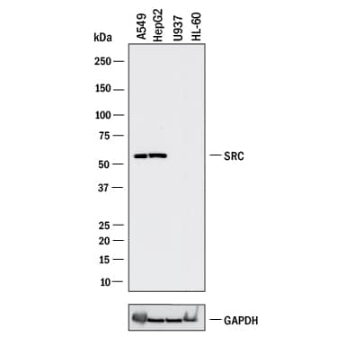
Western blot shows lysates of A549 human lung carcinoma cell line, HepG2 human hepatocellular carcinoma cell line, U937 human histiocytic lymphoma cell line (negative control), and HL-60 human acute promyelocytic leukemia cell line (negative control). PVDF membrane was probed with 2 µg/mL of Mouse Anti-Human Src Monoclonal Antibody (Catalog # MAB3807) followed by HRP-conjugated Anti-Mouse IgG Secondary Antibody (HAF018). A specific band was detected for Src at approximately 60 kDa (as indicated). GAPDH (MAB5718) is shown as a loading control. This experiment was conducted under reducing conditions and using Western Blot Buffer Group 1.Detection of Human and Mouse Src by Western Blot.
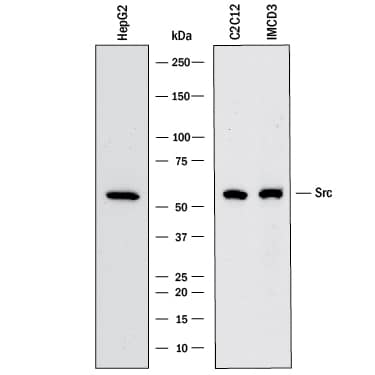
Western blot shows lysates of HepG2 human hepatocellular carcinoma cell line, C2C12 mouse myoblast cell line, and mIMCD-3 mouse epithelial cell line. PVDF membrane was probed with 2 µg/mL of Mouse Anti-Human Src Monoclonal Antibody (Catalog # MAB3807) followed by HRP-conjugated Anti-Mouse IgG Secondary Antibody (HAF018). A specific band was detected for Src at approximately 60 kDa (as indicated). This experiment was conducted under reducing conditions and using Western Blot Buffer Group 1.Src in Human Lung Cancer Tissue.
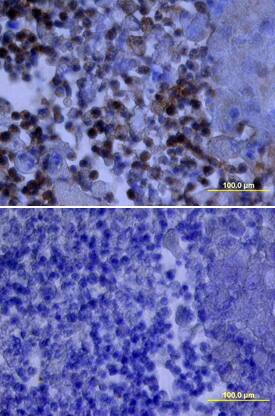
Src was detected in immersion fixed paraffin-embedded sections of human lung cancer tissue using Mouse Anti-Human Src Monoclonal Antibody (Catalog # MAB3807) at 25 µg/mL overnight at 4 °C. Tissue was stained using the Anti-Mouse HRP-DAB Cell & Tissue Staining Kit (brown; Catalog # CTS002) and counterstained with hematoxylin (blue). Lower panel shows a lack of labeling if primary antibodies are omitted and tissue is stained only with secondary antibody followed by incubation with detection reagents. View our protocol for Chromogenic IHC Staining of Paraffin-embedded Tissue Sections.Applications for Human Src Antibody
Immunohistochemistry
Sample: Immersion fixed paraffin-embedded sections of human liver cancer tissue and human lung cancer tissue
Western Blot
Sample: A549 human lung carcinoma cell line, HepG2 human hepatocellular carcinoma cell line, C2C12 mouse myoblast cell line, and mIMCD‑3 mouse epithelial cell line
Formulation, Preparation, and Storage
Purification
Reconstitution
Formulation
Shipping
Stability & Storage
- 12 months from date of receipt, -20 to -70 °C as supplied.
- 1 month, 2 to 8 °C under sterile conditions after reconstitution.
- 6 months, -20 to -70 °C under sterile conditions after reconstitution.
Background: Src
The Src family of proteins are intracellular tyrosine kinases involved in cell proliferation, differentiation, motility, and survival. Src family activity is regulated by tyrosine phosphorylation at two sites with opposing effects. Autophosphorylation in the activation loop of the kinase domain (Y419 of human Src) up‑regulates the enzyme, while phosphorylation in the C-terminal tail (Y530 of human Src) down‑regulates activity.
Long Name
Alternate Names
Gene Symbol
UniProt
Additional Src Products
Product Documents for Human Src Antibody
Product Specific Notices for Human Src Antibody
For research use only


