Human STAT1 Antibody
R&D Systems, part of Bio-Techne | Catalog # MAB14901

Key Product Details
Validated by
Species Reactivity
Validated:
Cited:
Applications
Validated:
Cited:
Label
Antibody Source
Product Specifications
Immunogen
Ala687-Val750
Accession # P42224
Specificity
Clonality
Host
Isotype
Scientific Data Images for Human STAT1 Antibody
Detection of Human STAT1 by Western Blot.
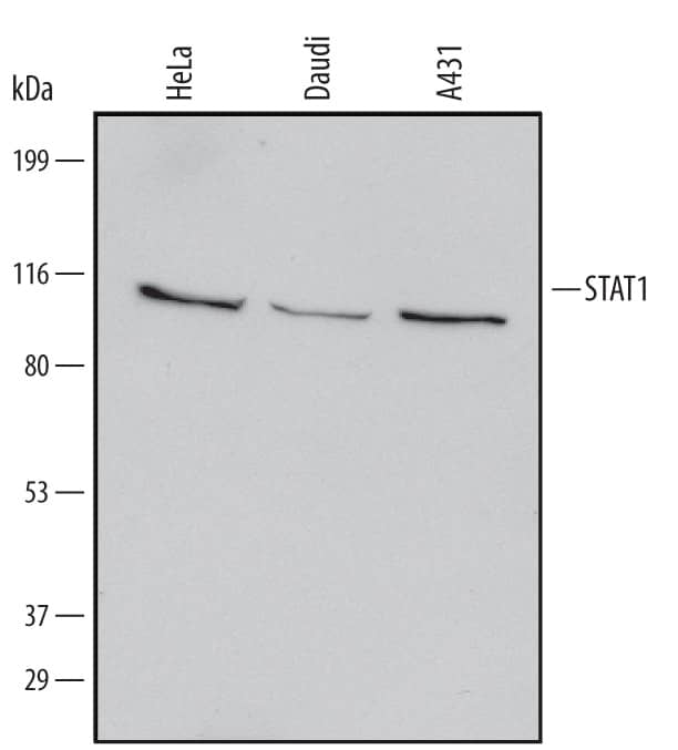
Western blot shows lysates of HeLa human cervical epithelial carcinoma cell line, Daudi human Burkitt's lymphoma cell line, and A431 human epithelial carcinoma cell line. PVDF Membrane was probed with 1 µg/mL of Mouse STAT1 Monoclonal Antibody (Catalog # MAB14091) followed by HRP-conjugated Anti-Mouse IgG Secondary Antibody (Catalog # HAF007). A specific band was detected for STAT1 at approximately 90 kDa (as indicated). This experiment was conducted under reducing conditions and using Immunoblot Buffer Group 1.Western Blot Shows Human STAT1 Specificity by Using Knockout Cell Line.
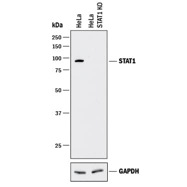
Western blot shows lysates of HeLa human cervical epithelial carcinoma parental cell line and STAT1 knockout HeLa cell line (KO). PVDF membrane was probed with 1 µg/mL of Mouse Anti-Human STAT1 Monoclonal Antibody (Catalog # MAB14901) followed by HRP-conjugated Anti-Mouse IgG Secondary Antibody (Catalog # HAF018). A specific band was detected for STAT1 at approximately 90 kDa (as indicated) in the parental HeLa cell line, but is not detectable in knockout HeLa cell line. GAPDH (Catalog # MAB5718) is shown as a loading control. This experiment was conducted under reducing conditions and using Immunoblot Buffer Group 1.STAT1 Specificity is Shown by Immunocytochemistry in Knockout Cell Line.
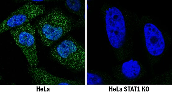
STAT1 was detected in immersion fixed HeLa human cervical epithelial carcinoma cell line treated with IFN-alpha 1 but is not detected in STAT1 knockout (KO) HeLa cell line using Mouse Anti-Human STAT1 Monoclonal Antibody (Catalog # MAB14901) at 1 µg/mL for 3 hours at room temperature. Cells were stained using the NorthernLights™ 493-conjugated Anti-Mouse IgG Secondary Antibody (green; Catalog # NL009) and counterstained with DAPI (blue). Specific staining was localized to cytoplasm and nuclei. View our protocol for Fluorescent ICC Staining of Cells on Coverslips.Applications for Human STAT1 Antibody
Knockout Validated
Western Blot
Sample: HeLa human cervical epithelial carcinoma cell line, Daudi human Burkitt's lymphoma cell line, and A431 human epithelial carcinoma cell line
Formulation, Preparation, and Storage
Purification
Reconstitution
Formulation
Shipping
Stability & Storage
- 12 months from date of receipt, -20 to -70 °C as supplied.
- 1 month, 2 to 8 °C under sterile conditions after reconstitution.
- 6 months, -20 to -70 °C under sterile conditions after reconstitution.
Background: STAT1
STAT1 is a member of the STAT family of cytoplasmic transcription factors that mediate cytokine, growth factor and hormone receptor signal transduction. STAT1 is associated with type I and II interferon signaling. Phosphorylation of STAT1a at Y701 leads to dimerization and translocation to the nucleus to activate gene transcription. Human STAT1 shows 93% and 94% aa identity with mouse and rat STAT1, respectively, over the region used as an immunogen. This region is identical between isoforms STAT1a (91 kDa) and STAT1b (84 kDa).
Long Name
Alternate Names
Gene Symbol
UniProt
Additional STAT1 Products
Product Documents for Human STAT1 Antibody
Product Specific Notices for Human STAT1 Antibody
For research use only


