Human TAFA2/FAM19A2 Antibody
R&D Systems, part of Bio-Techne | Catalog # AF4179

Key Product Details
Species Reactivity
Applications
Label
Antibody Source
Product Specifications
Immunogen
Ala31-His131
Accession # Q8N3H0
Specificity
Clonality
Host
Isotype
Endotoxin Level
Scientific Data Images for Human TAFA2/FAM19A2 Antibody
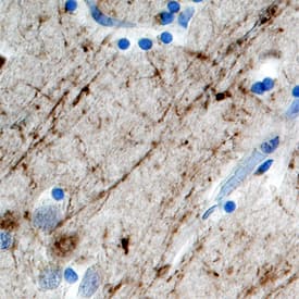
TAFA2/FAM19A2 in Human Brain.
TAFA2/FAM19A2 was detected in immersion fixed paraffin-embedded sections of human brain (cortex) using Human TAFA2/FAM19A2 Antigen Affinity-purified Polyclonal Antibody (Catalog # AF4179) at 10 µg/mL overnight at 4 °C. Before incubation with the primary antibody, tissue was subjected to heat-induced epitope retrieval using Antigen Retrieval Reagent-Basic (Catalog # CTS013). Tissue was stained using the Anti-Sheep HRP-DAB Cell & Tissue Staining Kit (brown; Catalog # CTS019) and counterstained with hematoxylin (blue). Specific staining was localized to neuronal processes. View our protocol for Chromogenic IHC Staining of Paraffin-embedded Tissue Sections.Applications for Human TAFA2/FAM19A2 Antibody
Immunohistochemistry
Sample: Immersion fixed paraffin-embedded sections of human brain (cortex)
Neutralization
Formulation, Preparation, and Storage
Purification
Reconstitution
Formulation
Shipping
Stability & Storage
- 12 months from date of receipt, -20 to -70 °C as supplied.
- 1 month, 2 to 8 °C under sterile conditions after reconstitution.
- 6 months, -20 to -70 °C under sterile conditions after reconstitution.
Background: TAFA2/FAM19A2
TAFA2 (also FAM19A2) is a secreted, 11 kDa member of the FAM19/TAFA family of chemokine-like proteins (1). It is synthesized as a 131 amino acid (aa) precursor that contains a 30 aa signal sequence and a 101 aa mature chain. Like other members of the FAM19/TAFA family, with the exception of TAFA5, mature TAFA1 contains 10 regularly spaced cysteine residues that follow the pattern CX7CCX13CXCX14CX11CX4CX5CX10C, where C represents a conserved cysteine residue and X represents any noncysteine amino acid (1). Human TAFA2 is 97% aa identical to mouse TAFA2 (1). TAFA2 expression can be detected in the central nervous system (CNS), colon, heart, lung, spleen, kidney, and thymus, but its expression in the CNS is 50- to 1000-fold higher than in other tissues (1). Within the CNS, TAFA2 expression is highest in the occipital and frontal cortex (3- to 10-fold more abundantly expressed than in other cortical regions) and medulla (1). The biological functions of TAFA family members remain to be determined, but there are a few tentative hypotheses. First, TAFAs may modulate immune responses in the CNS by functioning as brain-specific chemokines, and may act with other chemokines to optimize the recruitment and activity of immune cells in the CNS (1). Second, TAFAs may represent a novel class of neurokines that act as regulators of immune nervous cells (1 - 2). Finally, TAFAs may control axonal sprouting following brain injury (1).
References
- Tang, Y.T. et al. (2004) Genomics 83:727.
- Benveniste, E. (1998) Cytokine Growth Factor Rev. 9:259.
Long Name
Alternate Names
Gene Symbol
UniProt
Additional TAFA2/FAM19A2 Products
Product Documents for Human TAFA2/FAM19A2 Antibody
Product Specific Notices for Human TAFA2/FAM19A2 Antibody
For research use only
