Human TMED1 Antibody
R&D Systems, part of Bio-Techne | Catalog # MAB2243


Conjugate
Catalog #
Key Product Details
Species Reactivity
Human
Applications
CyTOF-ready, Flow Cytometry, Immunocytochemistry
Label
Unconjugated
Antibody Source
Monoclonal Mouse IgG1 Clone # 1009527
Product Specifications
Immunogen
Human embryonic kidney cell, HEK293 derived human TMED1
Met1-Asn194
Accession # Q13445
Met1-Asn194
Accession # Q13445
Specificity
Detects human TMED1 in direct ELISAs.
Clonality
Monoclonal
Host
Mouse
Isotype
IgG1
Scientific Data Images for Human TMED1 Antibody
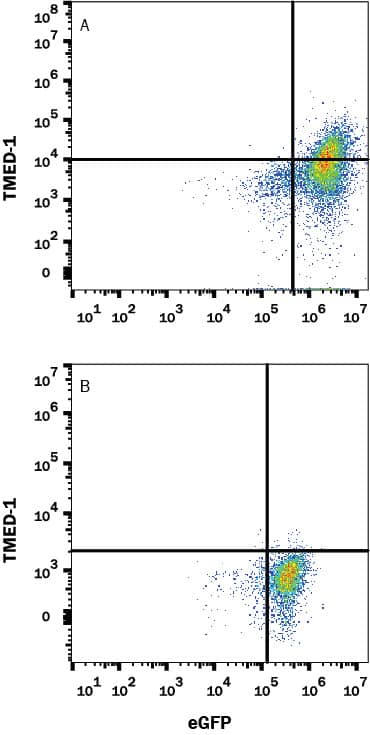
Detection of TMED-1 in HEK293 Human Cell Line Transfected with Human TMED-1 and eGFP by Flow Cytometry.
HEK293 human embryonic kidney cell line transfected with (A) TMED-1 or (B) irrelevant protein, and eGFP were stained with Mouse Anti-Human TMED-1 Monoclonal Antibody (Catalog # MAB2243) followed by Allophycocyanin-conjugated Anti-Mouse IgG Secondary Antibody (Catalog # F0101B). Quadrant markers were set based Mouse IgG1 Isotype Control Antibody staining (Catalog # MAB002, data not shown). View our protocol for Staining Membrane-associated Proteins.TMED1 in A549 Human Cell Line.
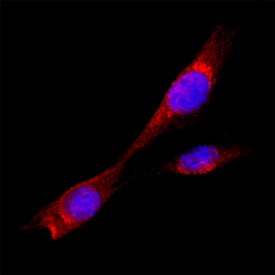
TMED1 was detected in immersion fixed A549 human lung carcinoma cell line using Mouse Anti-Human TMED1 Monoclonal Antibody (Catalog # MAB2243) at 8 µg/mL for 3 hours at room temperature. Cells were stained using the NorthernLights™ 557-conjugated Anti-Mouse IgG Secondary Antibody (red; Catalog # NL007) and counterstained with DAPI (blue). Specific staining was localized to cytoplasm. View our protocol for Fluorescent ICC Staining of Cells on Coverslips.Applications for Human TMED1 Antibody
Application
Recommended Usage
CyTOF-ready
Ready to be labeled using established conjugation methods. No BSA or other carrier proteins that could interfere with conjugation.
Flow Cytometry
0.25 µg/106 cells
Sample: HEK293 Human Cell Line Transfected with Human TMED-1 and eGFP
Sample: HEK293 Human Cell Line Transfected with Human TMED-1 and eGFP
Immunocytochemistry
8-25 µg/mL
Sample: Immersion fixed A549 human lung carcinoma cell line
Sample: Immersion fixed A549 human lung carcinoma cell line
Formulation, Preparation, and Storage
Purification
Protein A or G purified from hybridoma culture supernatant
Reconstitution
Reconstitute at 0.5 mg/mL in sterile PBS. For liquid material, refer to CoA for concentration.
Formulation
Lyophilized from a 0.2 μm filtered solution in PBS with Trehalose. *Small pack size (SP) is supplied either lyophilized or as a 0.2 µm filtered solution in PBS.
Shipping
Lyophilized product is shipped at ambient temperature. Liquid small pack size (-SP) is shipped with polar packs. Upon receipt, store immediately at the temperature recommended below.
Stability & Storage
Use a manual defrost freezer and avoid repeated freeze-thaw cycles.
- 12 months from date of receipt, -20 to -70 °C as supplied.
- 1 month, 2 to 8 °C under sterile conditions after reconstitution.
- 6 months, -20 to -70 °C under sterile conditions after reconstitution.
Background: TMED1
References
- Connolly, D. et al. (2013) J Biol Chem. 288:5616.
- Gour, N. and Lajoie, S. (2018) Curr Allergy Ashma Rep. 16:65.
- Jenne, N. (2002) J Biol Chem. 277:46504.
- Anantharaman, V. and Aravind, L. (2002) Genome Biol. 3:research0023
- Gomez-Navarro, N. and Miller, E. (2016) J Cell Biol. 215:769.
- Hardman, C. and Ogg, G. (2016). Curr Opin Immunol. 42:16.
Alternate Names
IL-1RL1LG, IL1RL1-Binding Protein, Il1rl1l, IL1RL1LG, Interleukin 1 Receptor-Like 1 Ligand, Ly84l, P24g1, P24gamma1, Tp24
Gene Symbol
TMED1
UniProt
Additional TMED1 Products
Product Documents for Human TMED1 Antibody
Product Specific Notices for Human TMED1 Antibody
For research use only
Loading...
Loading...
Loading...
Loading...
Loading...
