Human TNF-alpha Antibody
R&D Systems, part of Bio-Techne | Catalog # MAB610R
Recombinant Monoclonal Antibody.

Key Product Details
Species Reactivity
Human
Applications
ELISA Capture (Matched Antibody Pair), Neutralization
Label
Unconjugated
Antibody Source
Recombinant Monoclonal Mouse IgG1 Clone # 28401R
Product Specifications
Immunogen
E. coli-derived recombinant human TNF-alpha
Gly57-Leu233
Accession # P01375
Gly57-Leu233
Accession # P01375
Specificity
Detects human TNF-alpha in ELISAs and Western blots. In sandwich ELISAs, less than 0.05% cross-reactivity with recombinant human TNF‑ beta, recombinant mouse TNF‑ alpha, recombinant rat TNF‑ alpha, and recombinant porcine TNF‑ alpha is observed.
Clonality
Monoclonal
Host
Mouse
Isotype
IgG1
Endotoxin Level
<0.10 EU per 1 μg of the antibody by the LAL method.
Scientific Data Images for Human TNF-alpha Antibody
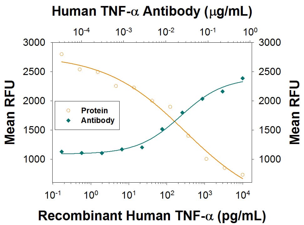
Cytotoxicity Induced by TNF-alpha and Neutralization by Human TNF-alpha Antibody.
Recombinant Human TNF-a (Catalog # 210-TA) induces cytotoxicity in the the L-929 mouse fibroblast cell line in a dose-dependent manner (orange line), as measured by Resazurin (Catalog # AR002). Cytotoxicity elicited by Recombinant Human TNF-a (0.25 ng/mL) is neutralized (green line) by increasing concentrations of Mouse Anti-Human TNF-a Recombinant Monoclonal Antibody (Catalog # MAB610R). The ND50 is typically 0.01-0.04 µg/mL in the presence of the metabolic inhibitor actinomycin D.Applications for Human TNF-alpha Antibody
Application
Recommended Usage
Neutralization
Measured by its ability to neutralize TNF-alpha -induced cytotoxicity in the L-929 mouse fibroblast cell line. Matthews, N. and M.L. Neale (1987) in Lymphokines and Interferons, A Practical Approach. Clemens, M.J. et al. (eds): IRL Press. 221. The Neutralization Dose (ND50) is typically 0.01-0.04 μg/mL in the presence of actinomycin D and 0.25 ng/mL Recombinant Human TNF-alpha.
Human TNF-alpha Sandwich Immunoassay
Please Note: Optimal dilutions of this antibody should be experimentally determined.
Formulation, Preparation, and Storage
Purification
Protein A or G purified from cell culture supernatant
Reconstitution
Reconstitute at 0.5
mg/mL in sterile PBS.
For liquid material, refer to CoA for concentration.
Formulation
Lyophilized from a 0.2 μm filtered solution in PBS with Trehalose. *Small pack size (SP) is supplied either lyophilized or as a 0.2 µm filtered solution in PBS.
Shipping
Lyophilized product is shipped at ambient temperature. Liquid small pack size (-SP) is shipped with polar packs. Upon receipt, store immediately at the temperature recommended below.
Stability & Storage
Use a manual defrost freezer and avoid repeated freeze-thaw cycles.
- 12 months from date of receipt, -20 to -70 °C as supplied.
- 1 month, 2 to 8 °C under sterile conditions after reconstitution.
- 6 months, -20 to -70 °C under sterile conditions after reconstitution.
Background: TNF-alpha
Long Name
Tumor Necrosis Factor alpha
Alternate Names
Cachetin, DIF, TNF, TNF-A, TNFA, TNFalpha, TNFG1F, TNFSF1A, TNFSF2
Entrez Gene IDs
Gene Symbol
TNF
UniProt
Additional TNF-alpha Products
Product Documents for Human TNF-alpha Antibody
Product Specific Notices for Human TNF-alpha Antibody
For research use only
Loading...
Loading...
Loading...
Loading...
