Human TNF RI/TNFRSF1A Antibody
R&D Systems, part of Bio-Techne | Catalog # MAB225


Conjugate
Catalog #
Key Product Details
Species Reactivity
Validated:
Human
Cited:
Human, Mouse, Rat
Applications
Validated:
CyTOF-ready, Flow Cytometry, Neutralization, Western Blot
Cited:
Cell Culture, Flow Cytometry, Immunocytochemistry/ Immunofluorescence, Immunohistochemistry, Neutralization, Western Blot
Label
Unconjugated
Antibody Source
Monoclonal Mouse IgG1 Clone # 16803
Product Specifications
Immunogen
E. coli-derived recombinant human TNF RI/TNFRSF1A
Accession # NP_001056
Accession # NP_001056
Specificity
Detects human TNF RI in Western blots. In Western blots, does not cross-react with recombinant human (rh) TNF RII, rmTNF RI, or rmTNF RII.
Clonality
Monoclonal
Host
Mouse
Isotype
IgG1
Endotoxin Level
<0.10 EU per 1 μg of the antibody by the LAL method.
Scientific Data Images for Human TNF RI/TNFRSF1A Antibody
TNF RI/TNFRSF1A Inhibition of TNF‑ alpha-induced Cytotoxicity and Neutralization by Human TNF RI/TNFRSF1A Antibody.
Recombinant Human TNF RI/TNFRSF1A (Catalog # 372-R1) inhibits Recombinant Human TNF-a (Catalog # 210-TA) induced cytotoxicity in the L-929 mouse fibroblast cell line in a dose-dependent manner (orange line), as measured by Resazurin (Catalog # AR002). Inhibition of Recombinant Human TNF-a (0.25 ng/mL) activity elicited by Recombinant Human TNF RI/TNFRSF1A (15 ng/mL) is neutralized (green line) by increasing concentrations of Mouse Anti-Human TNF RI/TNFRSF1A Monoclonal Antibody (Catalog # MAB225). The ND50 is typically 0.008-0.04 µg/mL in the presence of the metabolic inhibitor actinomycin D (1 µg/mL).Detection of TNF RI/TNFRSF1A in Human Blood Monocytes by Flow Cytometry.
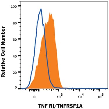
Human peripheral blood monocytes were stained with Mouse Anti-Human TNF RI/TNFRSF1A Monoclonal Antibody (Catalog # MAB225, filled histogram) or isotype control antibody (Catalog # MAB002, open histogram), followed by Allophycocyanin-conjugated Anti-Mouse IgG Secondary Antibody (Catalog # F0101B).Detection of Human TNF RI/TNFRSF1A by Immunohistochemistry
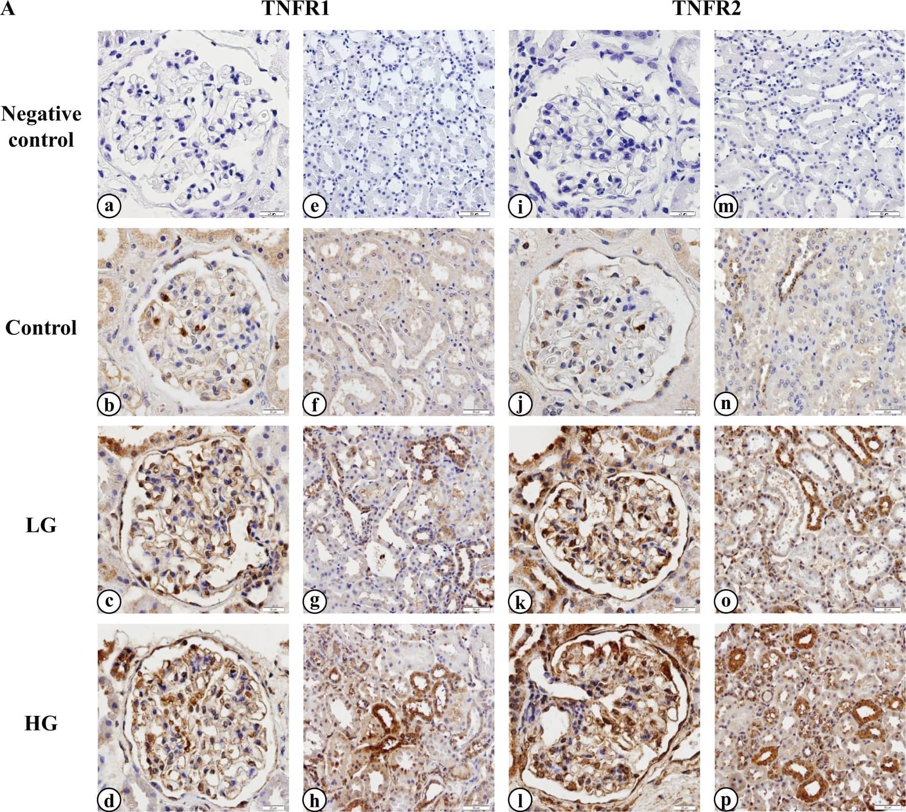
Representative immunohistochemical staining for TNFR1 and TNFR2 in the kidneys.(A) Images were captured at 200× (a, b, c, d, i, j, k, l) and 100× (e, f, g, h, m, n, o, p) magnification. Images show the negative controls in the glomeruli (a, i) and tubulointerstitium (e, m) and TNFR1 and TNFR2 immunostaining in the glomeruli (b, j) and tubulointerstitium (f, n) of the normal kidneys, respectively. TNFR1 and TNFR2 immunostaining is shown in the glomeruli (c, k) and tubulointerstitium (g, o) of the kidneys from selected IgAN patients who had levels of serum TNFR2 that ranked in the lowest 10 [low group (LG)]. TNFR1 and TNFR2 immunostaining in the glomeruli (d, l) and tubulointerstitium (h, p) of the kidneys from selected IgAN patients who had levels of serum TNFR2 that ranked in the highest 10 [high group (HG)], respectively, are also shown. (B) Percentage of the TNFR1 and TNFR2-positive area in the kidneys were evaluated. Glomerular and tubulointerstitial TNFR expression was elevated significantly in IgAN patients compared with those in control (Ctrl) subjects. The tubulointerstitial TNFR2-positive area was significantly larger in HG than in LG. However, there was no significant difference in the tubulointerstitial TNFR1 and glomerular TNFR areas between LG and HG. * P < 0.01, **P < 0.001, †P < 0.0001. Image collected and cropped by CiteAb from the following publication (https://dx.plos.org/10.1371/journal.pone.0122212), licensed under a CC-BY license. Not internally tested by R&D Systems.Applications for Human TNF RI/TNFRSF1A Antibody
Application
Recommended Usage
CyTOF-ready
Ready to be labeled using established conjugation methods. No BSA or other carrier proteins that could interfere with conjugation.
Flow Cytometry
0.25 µg/106 cells
Sample: Human peripheral blood monocytes
Sample: Human peripheral blood monocytes
Western Blot
1 µg/mL
Sample: Recombinant Human TNF RI/TNFRSF1A (Catalog # 636-R1)
under non-reducing conditions only
Sample: Recombinant Human TNF RI/TNFRSF1A (Catalog # 636-R1)
under non-reducing conditions only
Neutralization
Measured by its ability to neutralize TNF RI/TNFRSF1A-mediated inhibition of cytotoxicity in the L‑929 mouse fibroblast cell line. The Neutralization Dose (ND50) is typically 0.008-0.04 µg/mL in the presence of 15 ng/mL Recombinant Human TNF RI/TNFRSF1A, 0.25 ng/mL Recombinant Human TNF‑ alpha, and 1 µg/mL actinomycin D.
Formulation, Preparation, and Storage
Purification
Protein A or G purified from hybridoma culture supernatant
Reconstitution
Reconstitute at 0.5 mg/mL in sterile PBS. For liquid material, refer to CoA for concentration.
Formulation
Lyophilized from a 0.2 μm filtered solution in PBS with Trehalose. *Small pack size (SP) is supplied either lyophilized or as a 0.2 µm filtered solution in PBS.
Shipping
Lyophilized product is shipped at ambient temperature. Liquid small pack size (-SP) is shipped with polar packs. Upon receipt, store immediately at the temperature recommended below.
Stability & Storage
Use a manual defrost freezer and avoid repeated freeze-thaw cycles.
- 12 months from date of receipt, -20 to -70 °C as supplied.
- 1 month, 2 to 8 °C under sterile conditions after reconstitution.
- 6 months, -20 to -70 °C under sterile conditions after reconstitution.
Background: TNF RI/TNFRSF1A
Long Name
Tumor Necrosis Factor Receptor I
Alternate Names
CD120a, TNFRI, TNFRSF1A
Gene Symbol
TNFRSF1A
UniProt
Additional TNF RI/TNFRSF1A Products
Product Documents for Human TNF RI/TNFRSF1A Antibody
Product Specific Notices for Human TNF RI/TNFRSF1A Antibody
For research use only
Loading...
Loading...
Loading...
Loading...
Loading...

