Human TRAIL R2/TNFRSF10B Antibody
R&D Systems, part of Bio-Techne | Catalog # MAB631

Key Product Details
Validated by
Species Reactivity
Validated:
Cited:
Applications
Validated:
Cited:
Label
Antibody Source
Product Specifications
Immunogen
Met1-Glu182
Accession # CAG46696
Specificity
Clonality
Host
Isotype
Endotoxin Level
Scientific Data Images for Human TRAIL R2/TNFRSF10B Antibody
Human TRAIL R2/TNFRSF10B Antibody Induces Apoptosis of Jurkat Cells.
Human TRAIL R2/TNFRSF10B Monoclonal Antibody induces apoptosis of the Jurkat human acute T cell leukemia cell line in a dose-dependent manner, as measured by Resazurin (Catalog # AR002) . The ED50 for this effect is typically 2-12 ng/mL.Detection of Human TRAILR2/TNFRSF10B by Western Blot
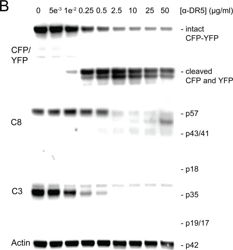
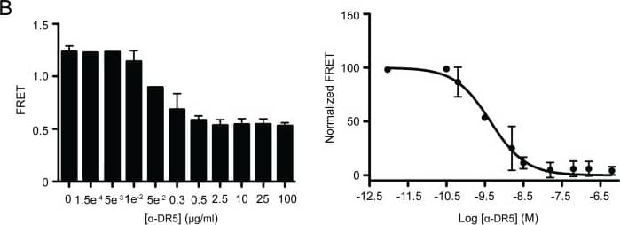
Cleavage of FRET probe is indicative of caspase activation.Cells were treated with (A) 0–300 ng/ml TRAIL for 2.5 hours or (B) 0–50 µg/ml anti-DR5 antibody for 20 h. The resultant cells were analyzed by western blotting using antibodies specific to caspase 3 (C3), caspase 8 (C8), and CFP/YFP. Image collected and cropped by CiteAb from the following publication (https://dx.plos.org/10.1371/journal.pone.0107010), licensed under a CC-BY license. Not internally tested by R&D Systems.Detection of Human TRAILR2/TNFRSF10B by Functional
MB231_CFP-YFP cells retain sensitivity to TRAIL and anti-DR5 antibody.(A) Parental MB231 (filled black) and MB231_CFP-YFP (open clear) cells were treated with TRAIL (0–100 ng/ml) for 2.5 h or anti-DR5 antibody (0–200 µg/ml) for 20 h, at the indicated concentrations. Cell viability was determined by MTT assay. (B) Cells were treated as in A and analyzed for apoptosis by flow cytometry after staining with Annexin-V-APC and propidium iodide (PI). p-values were determined using a Student’s t-test. EC50 values were determined using nonlinear curve fitting as described in the Materials and Methods section. For each EC50 value, 95% confidence intervals reflecting the statistical accuracy are reported in Table 1. Image collected and cropped by CiteAb from the following publication (https://dx.plos.org/10.1371/journal.pone.0107010), licensed under a CC-BY license. Not internally tested by R&D Systems.Applications for Human TRAIL R2/TNFRSF10B Antibody
Agonist Activity
Sample: Jurkat human acute T cell leukemia cell line
Formulation, Preparation, and Storage
Purification
Reconstitution
Formulation
Shipping
Stability & Storage
- 12 months from date of receipt, -20 to -70 °C as supplied.
- 1 month, 2 to 8 °C under sterile conditions after reconstitution.
- 6 months, -20 to -70 °C under sterile conditions after reconstitution.
Background: TRAIL R2/TNFRSF10B
Human TRAIL R2, also called DR5 and TRICK 2 is a type 1, TNF R family, membrane protein which is a receptor for TRAIL (APO2 ligand). In the new TNF superfamily nomenclature, TRAIL R2 is referred to as TNFRSF10B. TRAIL R2 cDNA encodes a 440 amino acid residue precursor protein containing extracellular cysteine-rich domains, a transmembrane domain and a cytoplasmic death domain. Among TNF receptor family proteins, TRAIL R2 is most closely related to TRAIL R1/DR4, sharing 55% amino acid sequence identity. Binding of trimeric TRAIL to TRAIL R2 induces apoptosis. The induction of apoptosis likely requires oligomerization of the receptor. The human TRAIL R2/Fc chimera neutralizes the ability of TRAIL to induce apoptosis. Besides TRAIL R2, an additional TRAIL R1/DR4, which tranduces apoptosis signaling, and two TRAIL decoy receptors, which antagonize TRAIL-induced apoptosis, have been reported.
References
- Chaudhary, P.M. et al. (1997) Immunity 7:821.
- Walczak, H. et al. (1997) EMBO J. 16:5386.
- Golstein, P. (1997) Curr. Biol. 7:R750.
Long Name
Alternate Names
Gene Symbol
UniProt
Additional TRAIL R2/TNFRSF10B Products
Product Documents for Human TRAIL R2/TNFRSF10B Antibody
Product Specific Notices for Human TRAIL R2/TNFRSF10B Antibody
For research use only


