Human TSC1 Antibody
R&D Systems, part of Bio-Techne | Catalog # MAB4379


Key Product Details
Species Reactivity
Validated:
Cited:
Applications
Validated:
Cited:
Label
Antibody Source
Product Specifications
Immunogen
Asp156-Thr300
Accession # Q92574
Specificity
Clonality
Host
Isotype
Scientific Data Images for Human TSC1 Antibody
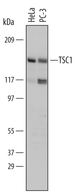
Detection of Human TSC1 by Western Blot.
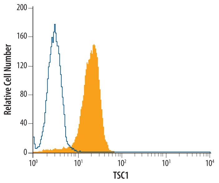
Western blot shows lysates of HeLa human cervical epithelial carcinoma cell line and PC-3 human prostate cancer cell line. PVDF Membrane was probed with 1 µg/mL of Human TSC1 Monoclonal Antibody (Catalog # MAB4379) followed by HRP-conjugated Anti-Mouse IgG Secondary Antibody (Catalog # HAF007). A specific band was detected for TSC1 at approximately 130 kDa (as indicated). This experiment was conducted under reducing conditions and using Immunoblot Buffer Group 1.Detection of TSC1 in Jurkat Human Cell Line by Flow Cytometry.
Jurkat human acute T cell leukemia cell line was stained with Human TSC1 Monoclonal Antibody (Catalog # MAB4379, filled histogram) or isotype control antibody (Catalog # MAB0041, open histogram), followed by Phycoerythrin-conjugated Anti-Mouse IgG F(ab')2Secondary Antibody (Catalog # F0102B). To facilitate intracellular staining, cells were fixed with PFA and permeabilized with saponin.Applications for Human TSC1 Antibody
CyTOF-ready
Intracellular Staining by Flow Cytometry
Sample: Jurkat human acute T cell leukemia cell line fixed with paraformaldehyde and permeabilized with saponin.
Western Blot
Sample: HeLa human cervical epithelial carcinoma cell line and PC‑3 human prostate cancer cell line
Formulation, Preparation, and Storage
Purification
Reconstitution
Formulation
Shipping
Stability & Storage
- 12 months from date of receipt, -20 to -70 °C as supplied.
- 1 month, 2 to 8 °C under sterile conditions after reconstitution.
- 6 months, -20 to -70 °C under sterile conditions after reconstitution.
Background: TSC1
TSC1 (Tuberous sclerosis 1), or hamartin, is a tumor suppressor which interacts with tumor suppressor TSC2 (tuberin) to form a cytoplasmic heterodimer. Mutations in either hamartin or tuberin are responsible for tuberous sclerosis (TSC), an autosomal dominant disease characterized by renal dysfunction, seizures, developmental delays, benign hamartomas and low grade neoplasms predominantly affecting the CNS, kidney, lung, skin, and heart. The TSC1/TSC2 complex suppresses cell growth by inhibiting mTOR, with TSC1 acting to inhibit the ubiquitination of TSC2, leading to increased cellular levels of TSC2 and thus enhancing its catalytic activity as a GTPase-activating protein for Rheb. TSC1 and TSC2 are also involved in the G2/M transition of the cell cycle through their interactions with CDK1 and cyclin B1. TSC1 has also been shown to interact with F-actin and ERM (Ezrin-Radixin-Moesin) proteins, implying a role in the modulation of cell adhesion and morphology.
Long Name
Alternate Names
Gene Symbol
UniProt
Additional TSC1 Products
Product Documents for Human TSC1 Antibody
Product Specific Notices for Human TSC1 Antibody
For research use only
