Human Tyro3/Dtk Antibody
R&D Systems, part of Bio-Techne | Catalog # AF859


Key Product Details
Species Reactivity
Validated:
Cited:
Applications
Validated:
Cited:
Label
Antibody Source
Product Specifications
Immunogen
Ala41-Ser428
Accession # Q06418
Specificity
Clonality
Host
Isotype
Scientific Data Images for Human Tyro3/Dtk Antibody
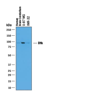
Detection of Human Tyro3/Dtk by Western Blot.
Western blot shows lysates of human brain (cerebellum) tissue, U-87 MG human glioblastoma/astrocytoma cell line, and IMR-32 human neuroblastoma cell line. PVDF membrane was probed with 1 µg/mL of Goat Anti-Human Tyro3/Dtk Antigen Affinity-purified Polyclonal Antibody (Catalog # AF859) followed by HRP-conjugated Anti-Goat IgG Secondary Antibody (Catalog # HAF019). A specific band was detected for Tyro3/Dtk at approximately 97 kDa (as indicated). This experiment was conducted under reducing conditions and using Immunoblot Buffer Group 1.Detection of Human Dtk/TYRO3 by Western Blot
rVSV∆G/EBOV entry kinetics: (A) VeroS cells were infected with rVSV∆G/EBOV at multiple MOIs, inoculum was removed after 1 h, and monitored for luciferase production over time. (B) VeroS, HEK293T, and HAP1 cells were infected with rVSV∆G/EBOV (MOI 1), inoculum was not removed, and monitored for luciferase production over time. Right panel zooms in to display the signals observed in HAP1 and HEK293T cells. (C) Immunoblots demonstrating production of the transfected receptors in HEK293T cells. (D) HEK293T cells transfected with the indicated receptors were infected with rVSV∆G/EBOV and monitored for luciferase production over time. Each experiment was repeated in duplicate, three independent times. Data shown are the averages of averages and corresponding SEM of three independent trials. Image collected and cropped by CiteAb from the following open publication (https://pubmed.ncbi.nlm.nih.gov/33348746), licensed under a CC-BY license. Not internally tested by R&D Systems.Applications for Human Tyro3/Dtk Antibody
Western Blot
Sample: Human brain (cerebellum) tissue, U‑87 MG human glioblastoma/astrocytoma cell line, and IMR‑32 human neuroblastoma cell line
Formulation, Preparation, and Storage
Purification
Reconstitution
Formulation
Shipping
Stability & Storage
- 12 months from date of receipt, -20 to -70 °C as supplied.
- 1 month, 2 to 8 °C under sterile conditions after reconstitution.
- 6 months, -20 to -70 °C under sterile conditions after reconstitution.
Background: Tyro3/Dtk
Axl (Ufo, Ark), Dtk (Sky, Tyro3, Rse, Brt) and Mer (human and mouse homologues of chicken c-Eyk) constitute a new receptor tyrosine kinase subfamily. The extracellular domain of these proteins contain two Ig-like motifs and two fibronectin type III motifs. This characteristic topology is also found in neural cell adhesion molecules and in receptor tyrosine phosphatases. All three receptors bind the vitamin K-dependent protein growth-arrest specific gene 6 (Gas6) which is structurally related to the anticoagulation factor protein S. The binding affinities for Gas6 is in the order of Axl > Dtk > Mer. Gas6 binding induces tyrosine phosphorylation and downstream signaling pathways that can lead to cell proliferation, migration, or the prevention of apoptosis. Dtk is widely expressed during embryonic development. In adults, Dtk is predominantly expressed in neurons in restricted regions of the brain.
References
- Nagata, K. et al. (1996) J. Biol. Chem. 22:30022.
- Crosier, K.E. and P.S Crosier (1997) Pathology 29:131.
Long Name
Alternate Names
Gene Symbol
UniProt
Additional Tyro3/Dtk Products
Product Documents for Human Tyro3/Dtk Antibody
Product Specific Notices for Human Tyro3/Dtk Antibody
For research use only
