Human uPAR Antibody
R&D Systems, part of Bio-Techne | Catalog # AF807


Key Product Details
Species Reactivity
Validated:
Cited:
Applications
Validated:
Cited:
Label
Antibody Source
Product Specifications
Immunogen
Leu23-Arg303
Accession # Q03405
Specificity
Clonality
Host
Isotype
Scientific Data Images for Human uPAR Antibody
Detection of Human uPAR by Western Blot.
Western blot shows lysates of A431 human epithelial carcinoma cell line and Saos-2 human osteosarcoma cell line. PVDF membrane was probed with 1 µg/mL of Goat Anti-Human uPAR Antigen Affinity-purified Polyclonal Antibody (Catalog # AF807) followed by HRP-conjugated Anti-Goat IgG Secondary Antibody (HAF017). Specific bands were detected for uPAR at approximately 30-60 kDa (as indicated). This experiment was conducted under reducing conditions and using Immunoblot Buffer Group 1.uPAR in Human Lung Cancer Tissue.
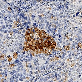
uPAR was detected in immersion fixed paraffin-embedded sections of human lung cancer tissue using Goat Anti-Human uPAR Antigen Affinity-purified Polyclonal Antibody (Catalog # AF807) at 1 µg/mL for 1 hour at room temperature followed by incubation with the Anti-Goat IgG VisUCyte™ HRP Polymer Antibody (VC004). Tissue was stained using DAB (brown) and counterstained with hematoxylin (blue). Specific staining was localized to cell membranes and cytoplasm. View our protocol for IHC Staining with VisUCyte HRP Polymer Detection Reagents.Detection of Human uPAR by Simple WesternTM.
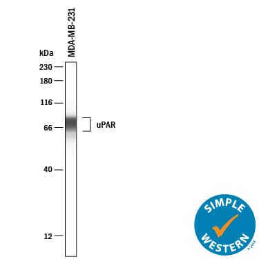
Simple Western lane view shows lysates of MDA-MB-231 human breast cancer cells, loaded at 0.2 mg/mL. A specific band was detected for uPAR at approximately 74 kDa (as indicated) using 20 µg/mL of Goat Anti-Human uPAR Antigen Affinity-purified Polyclonal Antibody (Catalog # AF807). This experiment was conducted under reducing conditions and using the 12-230kDa separation system.Applications for Human uPAR Antibody
Blockade of Receptor-ligand Interaction
Immunohistochemistry
Sample: Immersion fixed paraffin-embedded sections of human lung cancer tissue
Immunoprecipitation
Sample: U937 human histiocytic lymphoma cell line, see our available Western blot detection antibodies
Simple Western
Sample: MDA-MB-231 human breast cancer cells
Western Blot
Sample: A431 human epithelial carcinoma cell line and Saos‑2 human osteosarcoma cell line
Reviewed Applications
Read 1 review rated 4 using AF807 in the following applications:
Formulation, Preparation, and Storage
Purification
Reconstitution
Formulation
*Small pack size (-SP) is supplied either lyophilized or as a 0.2 µm filtered solution in PBS.
Shipping
Stability & Storage
- 12 months from date of receipt, -20 to -70 °C as supplied.
- 1 month, 2 to 8 °C under sterile conditions after reconstitution.
- 6 months, -20 to -70 °C under sterile conditions after reconstitution.
Background: uPAR
The urokinase-type Plasminogen Activator (uPA) is one of two activators that converts the extracellular zymogen plasminogen to plasmin, a serine protease that is involved in a variety of normal and pathological processes that require cell migration and/or tissue destruction. uPA is synthesized and released from cells as a single-chain (sc) pro-enzyme with limited enzymatic activity and is converted to an active two-chain (tc) disulfide-linked active enzyme by plasmin and other specific proteinases. Both the scuPA and tcuPA bind with high-affinity to the cell surface via the glycosyl phosphatidylinositol-linked receptor uPAR which serves to localize the uPA proteolytic activity. The enzymatic activity of scuPA has also been shown to be enhanced by binding to uPAR. Independent of their proteolytic activity, the uPA/uPAR interaction also initiates signal transduction responses resulting in activation of protein tyrosine kinases, gene expression, cell adhesion, and chemotaxis. uPAR can interact with integrins to suppress normal integrin adhesive function and promote adhesion to vitronectin through a high affinity vitronectin binding site on uPAR. uPAR cDNA encodes a 335 amino acid (aa) residue precursor protein with a 22 aa residue signal peptide, five potential N-linked glycosylation sites and a C‑terminal GPI-anchor site. An alternate spliced variant of uPAR encoding a secreted soluble form of uPAR also exists. Human and mouse uPAR share approximately 60% aa sequence identity and the receptor-ligand interaction is strictly species-specific.
References
- Dear, A.E. and R.L. Medcalf (1998) Eur. J. Biochemistry 252:185.
Long Name
Alternate Names
Gene Symbol
UniProt
Additional uPAR Products
Product Documents for Human uPAR Antibody
Product Specific Notices for Human uPAR Antibody
For research use only

