Human Wnt-7b Antibody
R&D Systems, part of Bio-Techne | Catalog # AF3460

Key Product Details
Species Reactivity
Validated:
Human
Cited:
Human, Mouse, Rat
Applications
Validated:
Immunohistochemistry, Western Blot
Cited:
Immunocytochemistry, Immunohistochemistry, Immunohistochemistry-Frozen, Immunohistochemistry-Paraffin, Western Blot
Label
Unconjugated
Antibody Source
Polyclonal Goat IgG
Product Specifications
Immunogen
E. coli-derived recombinant human Wnt-7b
Leu32-Glu72 & Thr216-Ala283
Accession # P56706
Leu32-Glu72 & Thr216-Ala283
Accession # P56706
Specificity
Detects human Wnt-7b in direct ELISAs and Western blots. In Western blots, approximately 20% cross-reactivity with recombinant human (rh) Wnt-7a is observed and less than 5% cross-reactivity with recombinant mouse (rm) Wnt-1, rmWnt-3a, rmWnt-4, rmWnt-5a, rhWnt-9a, rmWnt-11, and rmWnt-16 is observed.
Clonality
Polyclonal
Host
Goat
Isotype
IgG
Scientific Data Images for Human Wnt-7b Antibody
Detection of Human Wnt‑7b by Western Blot.
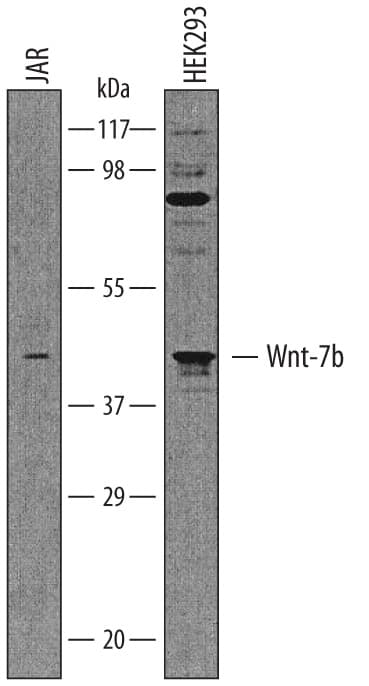
Western blot shows lysates of JAR human choriocarcinoma cell line and HEK293 human embryonic kidney cell line. PVDF membrane was probed with 1 µg/mL of Goat Anti-Human Wnt-7b Antigen Affinity-purified Polyclonal Antibody (Catalog # AF3460) followed by HRP-conjugated Anti-Goat IgG Secondary Antibody (HAF019). A specific band was detected for Wnt-7b at approximately 45 kDa (as indicated). This experiment was conducted under reducing conditions and using Immunoblot Buffer Group 1.Detection of Wnt-7b in Human Breast Cancer
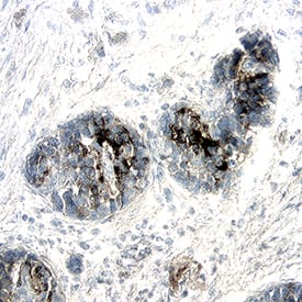
Wnt-7b was detected in immersion fixed paraffin-embedded sections of human breast cancer using Goat Anti-Human Wnt-7b Antigen Affinity-purified Polyclonal Antibody (Catalog # AF3460) at 15 µg/ml for 1 hour at room temperature followed by incubation with the Anti-Goat IgG VisUCyte™ HRP Polymer Antibody (Catalog # VC004). Before incubation with the primary antibody, tissue was subjected to heat-induced epitope retrieval using VisUCyte Antigen Retrieval Reagent-Basic (Catalog # VCTS021). Tissue was stained using DAB (brown) and counterstained with hematoxylin (blue). Specific staining was localized to cell surface and cytoplasm of cancer cells. View our protocol for IHC Staining with VisUCyte HRP Polymer Detection Reagents.Detection of Human Wnt-7b by Western Blot
Analysis of deltaNp73 in epidermal programming using an induced basal keratinocyte (iKC) model system.(A) Immunoblot of KLF4, p63 alpha, and p73 protein expression in neonatal human dermal fibroblast (HDFn) cells infected with lentivirus encoding deltaNp73 isoforms ( deltaNp73 alpha and deltaNp73 beta) or empty vector control in combination with KLF4 and deltaNp63 alpha. Cells were grown for 3 days and protein was harvested for immunoblot analysis. (B) Bar graphs of RNA expression for the indicated keratinocyte genes in HDFn cells infected in (A). Cells were grown for 3 days and RNA was harvested for qRT-PCR analysis. Expression data are represented as the fold increase relative to control. The mean of three replicates is shown with error bars representing SEM. *p-value < 0.05, **p-value < 0.01, ***p-value < 0.001. (C) Principal component analysis (PCA) plot of RNA-seq from HDFn cells infected with lentivirus encoding deltaNp73 beta or empty vector control in combination with KLF4 and deltaNp63 alpha. Cells were grown for 6 days and RNA was harvested for RNA-seq analysis. The percentage of variance contributed by each PC is listed in parentheses. (D and E) Tables listing the enriched Genome Ontology (GO) categories and pathways among the top 250 genes contributing to PC1 from (C). (F) Heatmap with the expression of a set of 44 genes that underlie the enrichment of GO categories from (D). Genes are annotated based on known roles in iKC-related processes (gray box) and the presence of a p63/p73 ChIP-seq peak within 50 kb of its TSS in multiple basal cell types (brown box). See also S5 and S6 Figs and S2–S9 Tables. Image collected and cropped by CiteAb from the following publication (https://pubmed.ncbi.nlm.nih.gov/31216312), licensed under a CC-BY license. Not internally tested by R&D Systems.Applications for Human Wnt-7b Antibody
Application
Recommended Usage
Immunohistochemistry
5-15 µg/mL
Sample: Immersion fixed paraffin-embedded sections of human pancreatic cancer tissue and human breast cancer tissue
Sample: Immersion fixed paraffin-embedded sections of human pancreatic cancer tissue and human breast cancer tissue
Western Blot
1 µg/mL
Sample: JAR human choriocarcinoma cell line and HEK293 human embryonic kidney cell line
Sample: JAR human choriocarcinoma cell line and HEK293 human embryonic kidney cell line
Formulation, Preparation, and Storage
Purification
Antigen Affinity-purified
Reconstitution
Reconstitute at 0.2 mg/mL in sterile PBS. For liquid material, refer to CoA for concentration.
Formulation
Lyophilized from a 0.2 μm filtered solution in PBS with Trehalose. *Small pack size (SP) is supplied either lyophilized or as a 0.2 µm filtered solution in PBS.
Shipping
Lyophilized product is shipped at ambient temperature. Liquid small pack size (-SP) is shipped with polar packs. Upon receipt, store immediately at the temperature recommended below.
Stability & Storage
Use a manual defrost freezer and avoid repeated freeze-thaw cycles.
- 12 months from date of receipt, -20 to -70 °C as supplied.
- 1 month, 2 to 8 °C under sterile conditions after reconstitution.
- 6 months, -20 to -70 °C under sterile conditions after reconstitution.
Background: Wnt-7b
Long Name
Wingless-type MMTV Integration Site Family, Member 7B
Alternate Names
Wnt7b
Gene Symbol
WNT7B
UniProt
Additional Wnt-7b Products
Product Documents for Human Wnt-7b Antibody
Product Specific Notices for Human Wnt-7b Antibody
For research use only
Loading...
Loading...
Loading...
Loading...


