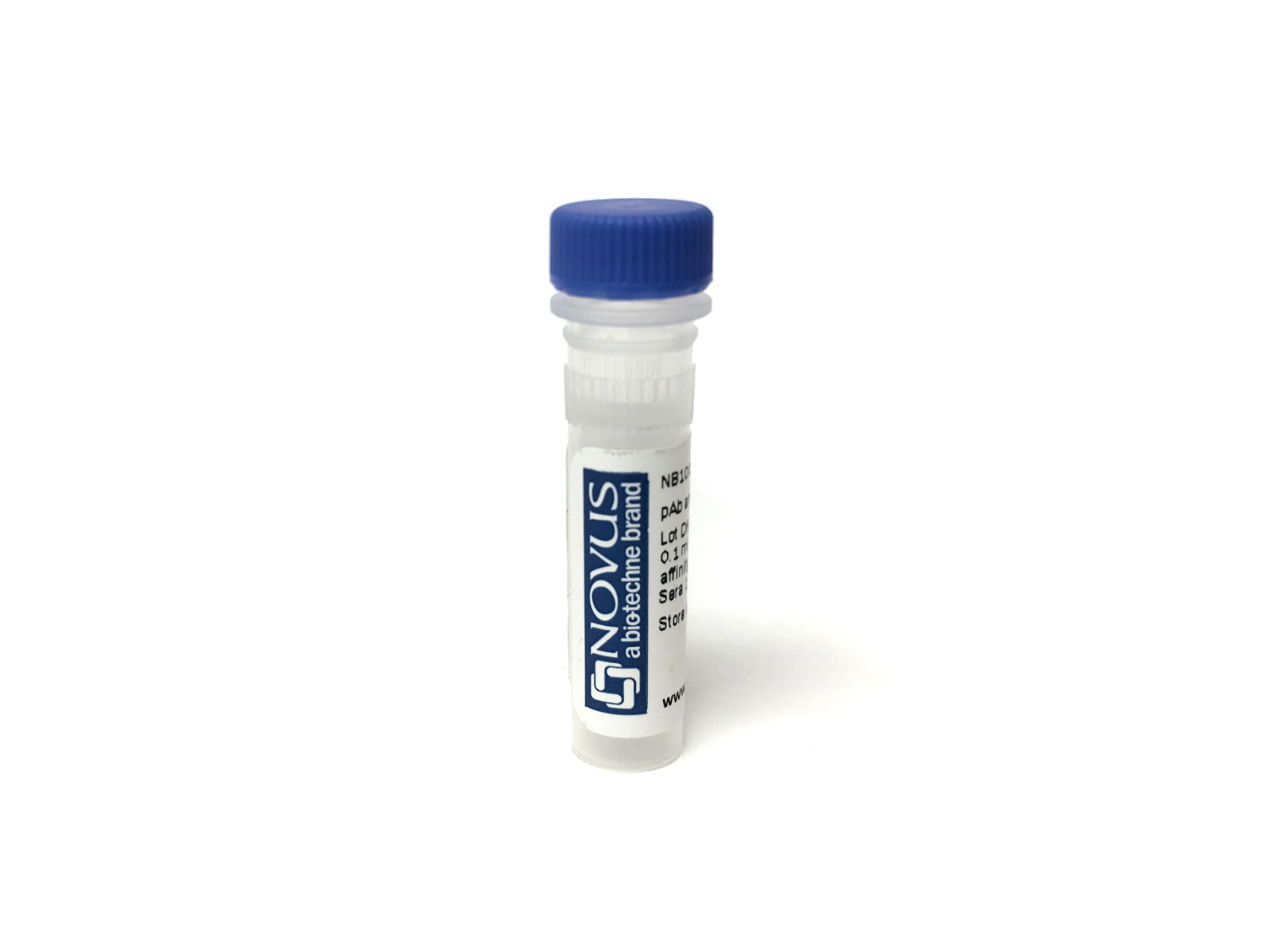
KLRG1 Antibody (1151A) [DyLight 488]
Novus Biologicals, part of Bio-Techne | Catalog # FAB6944K
Recombinant Monoclonal Antibody


Conjugate
Catalog #
Key Product Details
Species Reactivity
Mouse
Applications
Flow Cytometry
Label
DyLight 488 (Excitation = 493 nm, Emission = 518 nm)
Antibody Source
Recombinant Monoclonal Rabbit IgG Clone # 1151A
Concentration
Please see the vial label for concentration. If unlisted please contact technical services.
Product Specifications
Immunogen
Chinese hamster ovary cell line CHO-derived mouse KLRG1
Glu57-Tyr188
Accession # O88713
Glu57-Tyr188
Accession # O88713
Specificity
Detects mouse KLRG1 in flow cytometry.
Clonality
Monoclonal
Host
Rabbit
Isotype
IgG
Applications for KLRG1 Antibody (1151A) [DyLight 488]
Application
Recommended Usage
Flow Cytometry
Optimal dilutions of this antibody should be experimentally determined.
Application Notes
Optimal dilution of this antibody should be experimentally determined.
Formulation, Preparation, and Storage
Purification
Protein A or G purified from cell culture supernatant
Formulation
50mM Sodium Borate
Preservative
0.05% Sodium Azide
Concentration
Please see the vial label for concentration. If unlisted please contact technical services.
Shipping
The product is shipped with polar packs. Upon receipt, store it immediately at the temperature recommended below.
Stability & Storage
Store at 4C in the dark.
Background: KLRG1
KLRG1's primary ligand is E-cadherin, which is expressed on non-neural epithelial tissues, but the KLRG1 receptor also recognizes both N- and R-cadherin expressed in nervous system tissues (1,3,5,6). E-cadherin binding of KLRG1 leads to ITIM triggering and, ultimately, either reduced T cell proliferation or decreased NK cell cytotoxicity (3,5,6). More precisely, upon differentiation of CD8+ T cells, cells switch from signaling through CD28 and instead signal through the inhibitory KLRG1 receptor molecule (3,4). Phosphorylation of KLRG1's ITIM causes recruitment of two phosphatases, SH2-containing inositol polyphosphate 5-phoshate (SHIP-1) and SH-2 containing protein-tyrosine phosphatase 2 (SHP-2) (3,5). The effectors SHIP-1 and SHP-2 degrade PIP3 to PIP2, preventing Akt phosphorylation and inhibiting proliferation (3,5,6). Conversely, when KLRG1 signaling is blocked in highly differentiated T cells, Akt signaling is restored and cell proliferation resumes (3). An increase in highly differentiated T cells is observed in many age-related infections such as meningitis, pneumonia, and influenza, which also correlates with elevated KLRG1 levels (3). This observation suggests that KLRG1 may be a potential immunotherapeutic target, especially for vaccinations which typically have decreased response in aged patients (3).
References
1. Li Y, Hofmann M, Wang Q, Teng L, Chlewicki LK, Pircher H, Mariuzza RA. Structure of natural killer cell receptor KLRG1 bound to E-cadherin reveals basis for MHC-independent missing self recognition. Immunity. 2009 Jul 17;31(1):35-46. http://doi.org/10.1016/j.immuni.2009.04.019
2. Uniprot(Q96E93)
3. Henson SM, Akbar AN. KLRG1--more than a marker for T cell senescence. Age (Dordr). 2009 Dec;31(4):285-91. http://doi.org/10.1007/s11357-009-9100-9.
4. Thimme R, Appay V, Koschella M, Panther E, Roth E, Hislop AD, Rickinson AB, Rowland-Jones SL, Blum HE, Pircher H. Increased expression of the NK cell receptor KLRG1 by virus-specific CD8 T cells during persistent antigen stimulation. J Virol. 2005 Sep;79(18):12112-6.http://doi.org/10.1128/JVI.79.18.12112-12116.2005.
5. Borys SM, Bag AK, Brossay L, Adeegbe DO. The Yin and Yang of Targeting KLRG1+ Tregs and Effector Cells. Front Immunol. 2022 Apr 29;13:894508. http://doi.org10.3389/fimmu.2022.894508.
6. Van den Bossche J, Malissen B, Mantovani A, De Baetselier P, Van Ginderachter JA. Regulation and function of the E-cadherin/catenin complex in cells of the monocyte-macrophage lineage and DCs. Blood. 2012 Feb 16;119(7):1623-33. http://doi.org/10.1182/blood-2011-10-384289.
Long Name
Killer Cell Lectin-like Receptor Subfamily G Member 1
Alternate Names
CLEC15A, MAFA, MAFAL
Gene Symbol
KLRG1
Additional KLRG1 Products
Product Documents for KLRG1 Antibody (1151A) [DyLight 488]
Product Specific Notices for KLRG1 Antibody (1151A) [DyLight 488]
DyLight (R) is a trademark of Thermo Fisher Scientific Inc. and its subsidiaries.
This product is for research use only and is not approved for use in humans or in clinical diagnosis. Primary Antibodies are guaranteed for 1 year from date of receipt.
Loading...
Loading...
Loading...
Loading...
Loading...
Loading...