Laminin Antibody - BSA Free Best Seller
Novus Biologicals, part of Bio-Techne | Catalog # NB300-144


Conjugate
Catalog #
Key Product Details
Validated by
Biological Validation
Species Reactivity
Validated:
Human, Mouse, Rat, Chinese Hamster, Invertebrate, Mammal, Rabbit, Sheep
Cited:
Human, Mouse, Rat, Porcine, Avian - Chicken, Hamster - Cricetulus (Chinese Hamster), Invertebrate, Mammal, Ovine, Primate, Rabbit
Applications
Validated:
Flow Cytometry, Immunocytochemistry/ Immunofluorescence, Immunohistochemistry, Immunohistochemistry Free-Floating, Immunohistochemistry-Frozen, Immunohistochemistry-Paraffin, Western Blot
Cited:
Block/Neutralize, Flow Cytometry, IF/IHC, Immunocytochemistry, Immunocytochemistry/ Immunofluorescence, Immunohistochemistry, Immunohistochemistry-Frozen, Immunohistochemistry-Paraffin, Knockout Validated, Western Blot
Label
Unconjugated
Antibody Source
Polyclonal Rabbit IgG
Format
BSA Free
Concentration
1 mg/ml
Product Specifications
Immunogen
Laminin Antibody was made to Laminin 111 isolated from mouse Engelbreth-Holm-Swarm (EHS) sarcoma cells. [UniProt# P19137]
Reactivity Notes
Rabbit, Fruit Bat, Chinese Hamster, and S. mansoni reactivity reported in scientific literature (PMID: 18214989, 31877588, 29251349, and 28114363 respectively). Human, Mouse, Rat, and Sheep reported in multiple pieces of scientific literature.
Localization
Secreted, Basement membrane, Extracellular matrix
Specificity
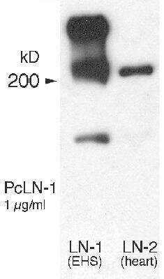
Laminin Antibody is pan-specific and reacts well with all Laminin isoforms tested: Laminin-1 (alpha-1, beta-1, and gamma-1) and Laminin-2 (alpha-2, beta-1, and gamma-1).
Marker
Basement Membrane Marker
Clonality
Polyclonal
Host
Rabbit
Isotype
IgG
Theoretical MW
337 kDa.
Disclaimer note: The observed molecular weight of the protein may vary from the listed predicted molecular weight due to post translational modifications, post translation cleavages, relative charges, and other experimental factors.
Disclaimer note: The observed molecular weight of the protein may vary from the listed predicted molecular weight due to post translational modifications, post translation cleavages, relative charges, and other experimental factors.
Scientific Data Images for Laminin Antibody - BSA Free
Staining of Laminin in Free Floating Rat Brain
Immunohistological analysis of brain stem section stained with rabbit polyclonal Laminin Antibody [NB300-144], dilution 1:1,000 in red, and costained with chicken pAb to Myelin Basic Protein (MBP), dilution 1:5,000 in green. The blue is DAPI staining of nuclear DNA. Following transcardial perfusion of rat with 4% paraformaldehyde, brain was post fixed for 24 hours, cut to 45 uM, and free-floating sections were stained with the above antibodies. The laminin antibody is an excellent marker of basement membranes surrounding blood vessels, while the MBP antibody stains the myelin sheathes around axons.Fluorescent Staining of Basement Membranes in Mouse Tissue Using Laminin Antibody
Laminin-Antibody-Immunohistochemistry-NB300-144-img0022.jpgStrong Staining of Laminin in Basement Membranes of Blood Vessels in Free Floating Mouse Tissue
Staining of mouse section of cortex stained with Laminin Antibody [NB300-144] (red). Blue is DAPI staining of DNA. This antibody reveals strong staining in the basement membranes of blood vessels.Applications for Laminin Antibody - BSA Free
Application
Recommended Usage
Immunocytochemistry/ Immunofluorescence
1:50-1:200
Immunohistochemistry
1:500-1:2000
Immunohistochemistry Free-Floating
1:1000-1:5000
Immunohistochemistry-Frozen
1:500-1:2000
Immunohistochemistry-Paraffin
1:500-1:2000
Western Blot
1:100-1:5000
Application Notes
This Laminin antibody detects bands at around 440, 220, and 158 kDa in Western Blot. Use in flow cytometry (PMID: 31819166) reported in scientific literature. Use in ICC/IF, IHC, IHC-Frozen, IHC-Paraffin, and Western Blot reported in multiple pieces of scientific literature. Immunostaining is enhanced by antigen retrieval with pepsin, especially paraffin tissue. br/>
The observed molecular weight of the protein may vary from the listed predicted molecular weight due to post translational modifications, post translation cleavages, relative charges, and other experimental factors.
The observed molecular weight of the protein may vary from the listed predicted molecular weight due to post translational modifications, post translation cleavages, relative charges, and other experimental factors.
Reviewed Applications
Read 9 reviews rated 4.6 using NB300-144 in the following applications:
Formulation, Preparation, and Storage
Purification
IgG purified
Formulation
50% PBS, 50% glycerol
Format
BSA Free
Preservative
0.035% Sodium Azide
Concentration
1 mg/ml
Shipping
The product is shipped with polar packs. Upon receipt, store it immediately at the temperature recommended below.
Stability & Storage
Aliquot and store at -20C or -80C. Avoid freeze-thaw cycles.
Background: Laminin
Laminins are an important and biologically active part of the basal lamina, influencing cell adhesion, differentiation, migration, signaling, neurite outgrowth and metastasis, where anti-laminin antibodies can be widely used to label blood vessels and basement membranes (1). Significant quantities of laminin are found in basement membranes, the thin extracellular matrices that surround epithelial tissue, nerve, fat cells and smooth, striated and cardiac muscle. Excessive serum laminin levels have been associated with fibrosis, cirrhosis and hepatitis, serious and frequent complications of chronic active liver disease characterized by excessive deposition of various normal components of connective tissue in liver (2). Epithelial mesenchymal transition (EMT) biomarkers include fibronectin, laminin, N-cadherin, and Slug (3).
References
1. Yang, M. Y., Chiao, M. T., Lee, H. T., Chen, C. M., Yang, Y. C., Shen, C. C., & Ma, H. I. (2015). An innovative three-dimensional gelatin foam culture system for improved study of glioblastoma stem cell behavior. J Biomed Mater Res B Appl Biomater, 103(3), 618-628. doi:10.1002/jbm.b.33214
2. Mak, K. M., & Mei, R. (2017). Basement Membrane Type IV Collagen and Laminin: An Overview of Their Biology and Value as Fibrosis Biomarkers of Liver Disease. Anat Rec (Hoboken), 300(8), 1371-1390. doi:10.1002/ar.23567
3. Choi, S., Yu, J., Park, A., Dubon, M. J., Do, J., Kim, Y., . . . Park, K. S. (2019). BMP-4 enhances epithelial mesenchymal transition and cancer stem cell properties of breast cancer cells via Notch signaling. Sci Rep, 9(1), 11724. doi:10.1038/s41598-019-48190-5
Alternate Names
LAMA, Laminin A chain, laminin subunit alpha-1, laminin, alpha 1, Laminin-1 subunit alpha, Laminin-3 subunit alpha, PTBHS, S-LAM alpha, S-laminin subunit alpha
Gene Symbol
LAMA1
UniProt
Additional Laminin Products
Product Documents for Laminin Antibody - BSA Free
Product Specific Notices for Laminin Antibody - BSA Free
This product is for research use only and is not approved for use in humans or in clinical diagnosis. Primary Antibodies are guaranteed for 1 year from date of receipt.
Loading...
Loading...
Loading...
Loading...
Loading...
Loading...


![Western Blot: Laminin Antibody [NB300-144] - Laminin Antibody](https://resources.bio-techne.com/images/products/nb300-144_rabbit-polyclonal-laminin-antibody-210202423454857.jpg)
![Western Blot: Laminin Antibody [NB300-144] - Laminin Antibody](https://resources.bio-techne.com/images/products/nb300-144_rabbit-polyclonal-laminin-antibody-210202423452412.jpg)
![Western Blot: Laminin Antibody [NB300-144] - Laminin Antibody](https://resources.bio-techne.com/images/products/nb300-144_rabbit-polyclonal-laminin-antibody-210202423454814.jpg)
![Western Blot: Laminin Antibody [NB300-144] - Laminin Antibody](https://resources.bio-techne.com/images/products/nb300-144_rabbit-polyclonal-laminin-antibody-210202423454825.jpg)
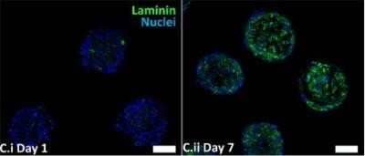
![Immunocytochemistry/ Immunofluorescence: Laminin Antibody [NB300-144] - Laminin Antibody](https://resources.bio-techne.com/images/products/nb300-144_rabbit-polyclonal-laminin-antibody-310202415171342.jpg)
![Western Blot: Laminin Antibody [NB300-144] - Laminin Antibody](https://resources.bio-techne.com/images/products/nb300-144_rabbit-polyclonal-laminin-antibody-310202416212338.jpg)