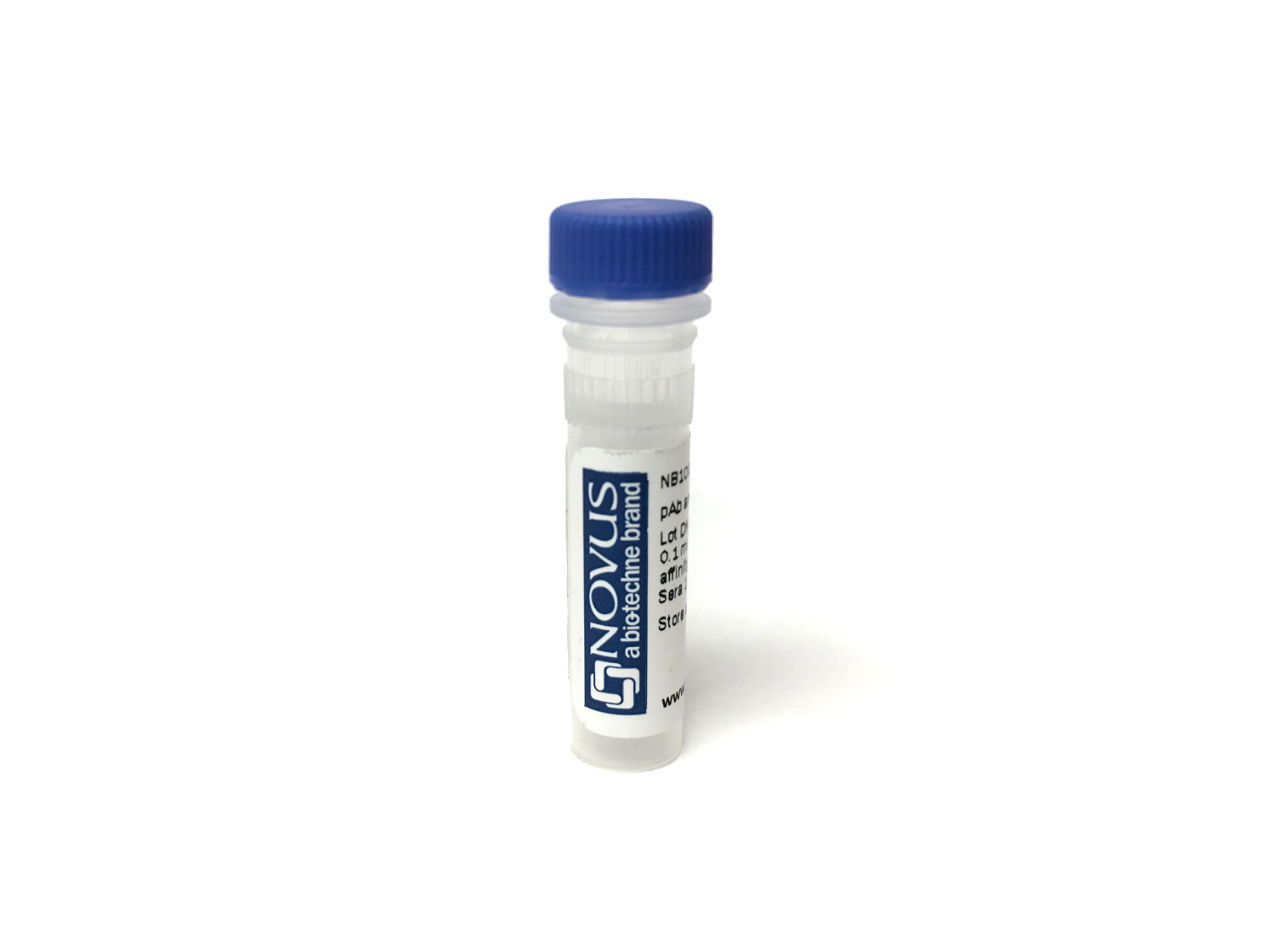
MAP2 Antibody (4H5) [mFluor Violet 450 SE]
Novus Biologicals, part of Bio-Techne | Catalog # NBP2-25156MFV450


Conjugate
Catalog #
Forumulation
Catalog #
Key Product Details
Species Reactivity
Human, Mouse, Rat, Bovine
Applications
Immunocytochemistry/ Immunofluorescence, Immunohistochemistry, Immunohistochemistry Free-Floating, Immunohistochemistry-Paraffin, Western Blot
Label
mFluor Violet 450 SE (Excitation = 406 nm, Emission = 445 nm)
Antibody Source
Monoclonal Mouse IgG1 Clone # 4H5
Concentration
Please see the vial label for concentration. If unlisted please contact technical services.
Product Specifications
Immunogen
MAP2 Antibody (4H5) was developed against full length purified bovine protein, epitope mapped to projection domain of human sequence, between amino acids 631 and 1056.
Localization
Cytoskeleton
Specificity
MAP2 Antibody (4H5) will be reactive to isoforms 1(MAP2B) and isoform 3 (MAP2A).
Marker
Neuronal Dendritic Marker
Clonality
Monoclonal
Host
Mouse
Isotype
IgG1
Applications for MAP2 Antibody (4H5) [mFluor Violet 450 SE]
Application
Recommended Usage
Immunocytochemistry/ Immunofluorescence
Optimal dilutions of this antibody should be experimentally determined.
Immunohistochemistry
Optimal dilutions of this antibody should be experimentally determined.
Immunohistochemistry Free-Floating
Optimal dilutions of this antibody should be experimentally determined.
Immunohistochemistry-Paraffin
Optimal dilutions of this antibody should be experimentally determined.
Western Blot
Optimal dilutions of this antibody should be experimentally determined.
Application Notes
Optimal dilution of this antibody should be experimentally determined.
Formulation, Preparation, and Storage
Purification
Immunogen affinity purified
Formulation
50mM Sodium Borate
Preservative
0.05% Sodium Azide
Concentration
Please see the vial label for concentration. If unlisted please contact technical services.
Shipping
The product is shipped with polar packs. Upon receipt, store it immediately at the temperature recommended below.
Stability & Storage
Store at 4C in the dark.
Background: MAP2
MAP2 isoforms are developmentally regulated and differentially expressed in neurons and some glia. MAP2c is predominantly expressed in the developing brain while the other isoforms are expressed in the adult brain. The distribution of MAP2 isoforms also varies, with MAP2a and MAP2b predominantly localized to dendrites, while MAP2c is also found in axons. Lastly, the expression of MAP2d is not limited to neurons and may be found in glia, specifically oligodendrocytes (1, 2). MAP2 isoforms associate with microtubules and mediate their interaction with actin filaments thereby playing a critical role in organizing the microtubule-actin network. In neurons, MAP2 isoforms are implicated in different processes including neurite initiation, elongation and stabilization as well as axon and dendrite formation (2). Knockout of MAP expression in animal models results in a variety of functional and structural brain defects according to the isoform affected (e.g., reduced LTP and LTD, reduced myelination, absence of corpus collosum, motor system malfunction, abnormal hippocampal dendritic morphology, abnormal synaptic plasticity) (4).
References
1. Dehmelt, L., & Halpain, S. (2005). The MAP2/Tau family of microtubule-associated proteins. Genome Biology. https://doi.org/10.1186/gb-2004-6-1-204
2. Mohan, R., & John, A. (2015). Microtubule-associated proteins as direct crosslinkers of actin filaments and microtubules. IUBMB Life. https://doi.org/10.1002/iub.1384
3. Shafit-Zagardo, B., & Kalcheva, N. (1998). Making sense of the multiple MAP-2 transcripts and their role in the neuron. Molecular Neurobiology. https://doi.org/10.1007/BF02740642
4. Tortosa, E., Kapitein, L. C., & Hoogenraad, C. C. (2016). Microtubule organization and microtubule-associated proteins (MAPs). In Dendrites: Development and Disease. https://doi.org/10.1007/978-4-431-56050-0_3
Long Name
Microtubule-associated Protein 2
Alternate Names
DKFZp686I2148, MAP-2, MAP2A, MAP2B, MAP2C, Microtubule Associated Protein 2, microtubule-associated protein 2, MTAP2
Gene Symbol
MAP2
Additional MAP2 Products
Product Documents for MAP2 Antibody (4H5) [mFluor Violet 450 SE]
Product Specific Notices for MAP2 Antibody (4H5) [mFluor Violet 450 SE]
mFluor(TM) is a trademark of AAT Bioquest, Inc. This conjugate is made on demand. Actual recovery may vary from the stated volume of this product. The volume will be greater than or equal to the unit size stated on the datasheet.
This product is for research use only and is not approved for use in humans or in clinical diagnosis. Primary Antibodies are guaranteed for 1 year from date of receipt.
Loading...
Loading...
Loading...
Loading...