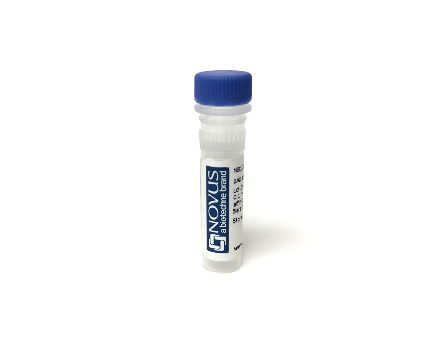
Melan-A/MART-1 Antibody (MLANA/7448R) - Azide and BSA Free
Novus Biologicals, part of Bio-Techne | Catalog # NBP3-21037
Recombinant Monoclonal Antibody


Conjugate
Catalog #
Key Product Details
Species Reactivity
Human
Applications
Immunohistochemistry-Paraffin
Label
Unconjugated
Antibody Source
Recombinant Monoclonal Rabbit IgG Kappa Clone # MLANA/7448R
Format
Azide and BSA Free
Concentration
1 mg/ml
Product Specifications
Immunogen
Recombinant fragment corresponding to the N-terminus of human Melan-A/MART-1 protein
Localization
Cytoplasm.
Specificity
This monoclonal antibody labels melanomas and other tumors showing melanocytic differentiation It is also a useful positive-marker for angiomyolipomas It does not stain tumor cells of epithelial, lymphoid, glial, or mesenchymal origin
Clonality
Monoclonal
Host
Rabbit
Isotype
IgG Kappa
Description
Positive Controls: COLO-38, SK-MEL-13 or SK-MEL-19 melanoma cell lines. Human skin or melanoma.
Antibody with azide - store at 2 to 8C. Antibody without azide - store at -20 to -80C. Non-hazardous. No MSDS required.
Antibody with azide - store at 2 to 8C. Antibody without azide - store at -20 to -80C. Non-hazardous. No MSDS required.
Applications for Melan-A/MART-1 Antibody (MLANA/7448R) - Azide and BSA Free
Application
Recommended Usage
Immunohistochemistry-Paraffin
1-2 ug/ml
Application Notes
Immunohistochemistry (Formalin-fixed): 1-2ug/ml for 30 minutes. at RT. Staining of formalin-fixed tissues requires heating tissue sections in 10mM Tris with 1mM EDTA, pH 9.0, for 45 min at 95C followed by cooling at RT for 20 minutes.
Optimal dilution for a specific application should be determined.
Optimal dilution for a specific application should be determined.
Formulation, Preparation, and Storage
Purification
Protein A or G purified
Formulation
10mM PBS
Format
Azide and BSA Free
Preservative
No Preservative
Concentration
1 mg/ml
Shipping
The product is shipped with polar packs. Upon receipt, store it immediately at the temperature recommended below.
Stability & Storage
Store at -20 to -70C. Avoid freeze-thaw cycles.
Background: Melan-A/MART-1
Alternate Names
Antigen LB39-AA, Antigen SK29-AA, MART-1, MelanA, MLANA
Gene Symbol
MLANA
Additional Melan-A/MART-1 Products
Product Documents for Melan-A/MART-1 Antibody (MLANA/7448R) - Azide and BSA Free
Product Specific Notices for Melan-A/MART-1 Antibody (MLANA/7448R) - Azide and BSA Free
This product is for research use only and is not approved for use in humans or in clinical diagnosis. Primary Antibodies are guaranteed for 1 year from date of receipt.
Loading...
Loading...
Loading...
Loading...