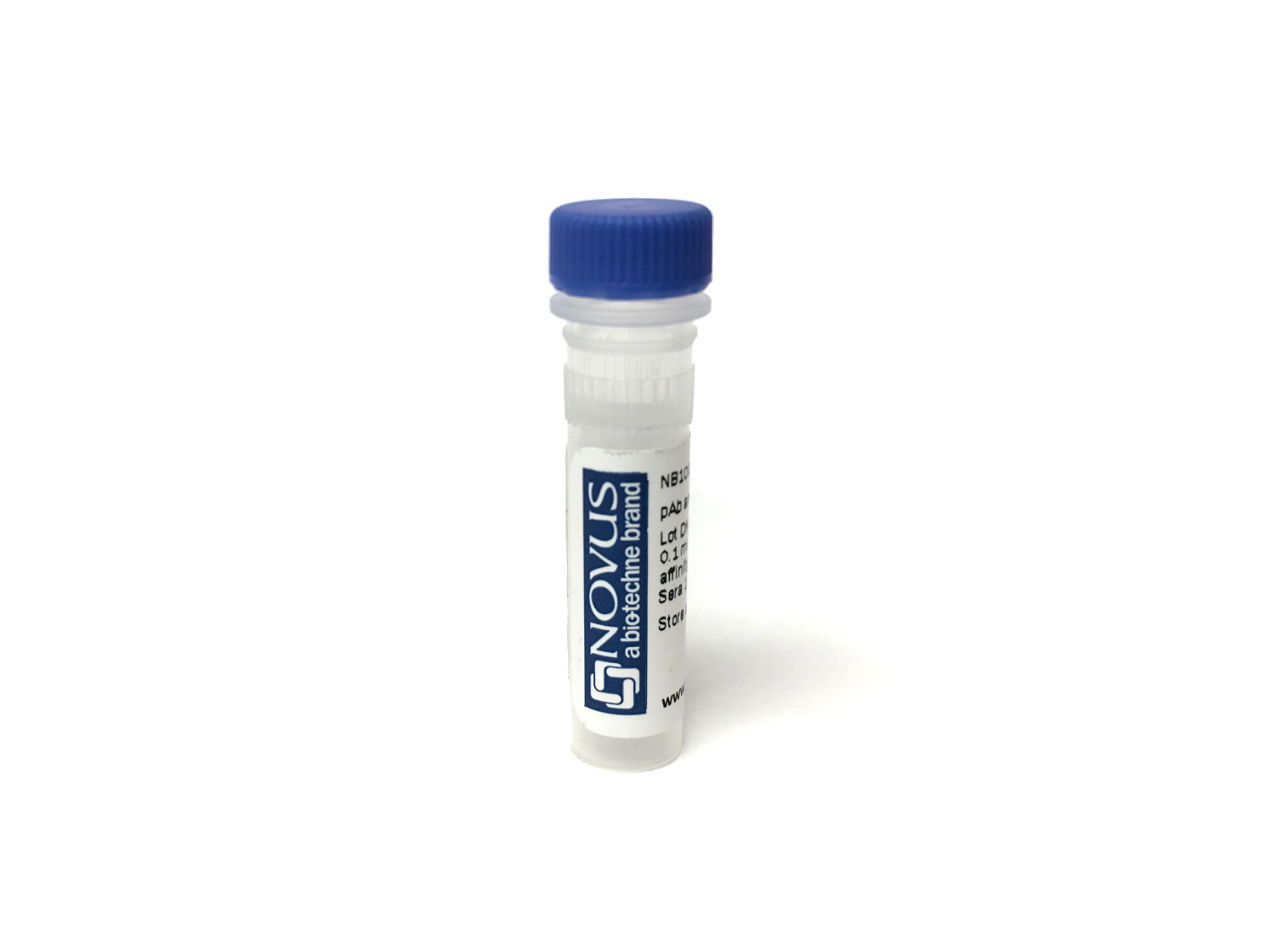
Melan-A/MART-1 Antibody (rMLANA/8180) [mFluor Violet 500 SE]
Novus Biologicals, part of Bio-Techne | Catalog # NBP3-24313MFV500
Recombinant Monoclonal Antibody


Conjugate
Catalog #
Forumulation
Catalog #
Key Product Details
Species Reactivity
Human
Applications
Immunohistochemistry-Paraffin
Label
mFluor Violet 500 SE (Excitation = 410 nm, Emission = 501 nm)
Antibody Source
Recombinant Monoclonal Mouse IgG1 kappa Clone # rMLANA/8180
Concentration
Please see the vial label for concentration. If unlisted please contact technical services.
Product Specifications
Immunogen
Recombinant full-length human Melan-A/MART-1 protein
Localization
Cytoplasm.
Specificity
This antibody recognizes a protein doublet of 20-22kDa, identified as MART-1 (Melanoma Antigen Recognized by T cells 1) or Melan-A. This monoclonal antibody labels melanomas and other tumors showing melanocytic differentiation. It is also a useful positive-marker for angiomyolipomas. It does not stain tumor cells of epithelial, lymphoid, glial, or mesenchymal origin.
Marker
Melanoma Marker
Clonality
Monoclonal
Host
Mouse
Isotype
IgG1 kappa
Applications for Melan-A/MART-1 Antibody (rMLANA/8180) [mFluor Violet 500 SE]
Application
Recommended Usage
Immunohistochemistry-Paraffin
Optimal dilutions of this antibody should be experimentally determined.
Application Notes
Optimal dilution of this antibody should be experimentally determined.
Formulation, Preparation, and Storage
Purification
Protein A or G purified
Formulation
50mM Sodium Borate
Preservative
0.05% Sodium Azide
Concentration
Please see the vial label for concentration. If unlisted please contact technical services.
Shipping
The product is shipped with polar packs. Upon receipt, store it immediately at the temperature recommended below.
Stability & Storage
Store at 4C in the dark.
Background: Melan-A/MART-1
Alternate Names
Antigen LB39-AA, Antigen SK29-AA, MART-1, MelanA, MLANA
Gene Symbol
MLANA
Additional Melan-A/MART-1 Products
Product Documents for Melan-A/MART-1 Antibody (rMLANA/8180) [mFluor Violet 500 SE]
Product Specific Notices for Melan-A/MART-1 Antibody (rMLANA/8180) [mFluor Violet 500 SE]
mFluor(TM) is a trademark of AAT Bioquest, Inc. This conjugate is made on demand. Actual recovery may vary from the stated volume of this product. The volume will be greater than or equal to the unit size stated on the datasheet.
This product is for research use only and is not approved for use in humans or in clinical diagnosis. Primary Antibodies are guaranteed for 1 year from date of receipt.
Loading...
Loading...
Loading...
Loading...