Mouse Alkaline Phosphatase/ALPL Antibody
R&D Systems, part of Bio-Techne | Catalog # AF2910

Key Product Details
Species Reactivity
Validated:
Cited:
Applications
Validated:
Cited:
Label
Antibody Source
Product Specifications
Immunogen
Phe18-Gly503
Accession # P09242
Specificity
Clonality
Host
Isotype
Scientific Data Images for Mouse Alkaline Phosphatase/ALPL Antibody
Detection of Mouse Alkaline Phosphatase/ALPL by Western Blot.
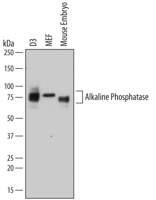
Western blot shows lysates of D3 mouse embryonic stem cell line, MEF mouse embryonic feeder cells, and mouse embryo tissue. PVDF membrane was probed with 1 µg/mL of Goat Anti-Mouse Alkaline Phosphatase/ALPL Antigen Affinity-purified Polyclonal Antibody (Catalog # AF2910) followed by HRP-conjugated Anti-Goat IgG Secondary Antibody (Catalog # HAF109). Specific bands were detected for Alkaline Phosphatase/ALPL at approximately 75-80 kDa (as indicated). This experiment was conducted under reducing conditions and using Immunoblot Buffer Group 1.Detection of ALPL in Rat Stem Cells by Flow Cytometry.
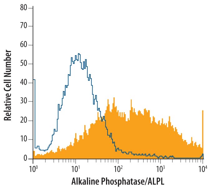
Rat Mesenchymal Stem Cells were stained with Goat Anti-Mouse Alkaline Phosphatase/ALPL Antigen Affinity-purified Polyclonal Antibody (Catalog # AF2910, filled histogram) or isotype control antibody (Catalog # AB-108-C, open histogram), followed by Phycoerythrin-conjugated Anti-Goat IgG Secondary Antibody (Catalog # F0107).Alkaline Phosphatase/ALPL in Rat Mesenchymal Stem Cells.
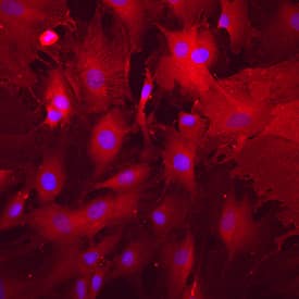
Alkaline Phosphatase/ALPL was detected in immersion fixed rat mesenchymal stem cells using Goat Anti-Mouse Alkaline Phosphatase/ALPL Antigen Affinity-purified Polyclonal Antibody (Catalog # AF2910) at 10 µg/mL for 3 hours at room temperature. Cells were stained using the NorthernLights™ 557-conjugated Anti-Goat IgG Secondary Antibody (red; Catalog # NL001) and counterstained with DAPI (blue). Specific staining was localized to cell surfaces and cytoplasm. View our protocol for Fluorescent ICC Staining of Cells on Coverslips.Applications for Mouse Alkaline Phosphatase/ALPL Antibody
CyTOF-ready
Flow Cytometry
Sample: Rat mesenchymal stem cells
Immunocytochemistry
Sample: Immersion fixed rat mesenchymal stem cells
Immunohistochemistry
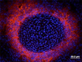
Sample: Immersion fixed frozen sections of mouse embryonic (E13.5) developing vertebra
Immunoprecipitation
Sample: Conditioned cell culture medium spiked with Recombinant Mouse Alkaline Phosphatase/ALPL (Catalog # 2910-AP), see our available Western blot detection antibodies
Western Blot
Sample: D3 mouse embryonic stem cell line, MEF mouse embryonic feeder cells, and mouse embryo tissue
Reviewed Applications
Read 3 reviews rated 5 using AF2910 in the following applications:
Formulation, Preparation, and Storage
Purification
Reconstitution
Formulation
Shipping
Stability & Storage
- 12 months from date of receipt, -20 to -70 °C as supplied.
- 1 month, 2 to 8 °C under sterile conditions after reconstitution.
- 6 months, -20 to -70 °C under sterile conditions after reconstitution.
Background: Alkaline Phosphatase/ALPL
Several distinct genes encode alkaline phosphatases (APs) in mice with different tissue-specific expression patterns. The Alpl gene, also known as Akp2, encodes the liver/bone/kidney isozyme, also known as the tissue-nonspecific AP (TNAP) (1). The Alpl gene is a key regulator of bone mineralization in mice (2). A variety of mutations in the human ALPL gene leads to different forms of hypophosphatasia, characterized by poorly mineralized cartilage and bones (3). The native ALPL is a glycosylated homodimer attached to the membrane through a GPI-anchor. The C-terminal pro peptide (residues 504 to 524) is not present in the mature form.
References
- Terao, M. and B. Mintz (1987) Proc. Natl. Acad. Sci. USA 84:7051.
- Hessle, L. et al. (2002) Proc. Natl. Acad. Sci. USA 99:9445.
- Di Mauro, S. et al. (2002) J. Bone Miner. Res. 17:1383.
Long Name
Alternate Names
Gene Symbol
UniProt
Additional Alkaline Phosphatase/ALPL Products
Product Documents for Mouse Alkaline Phosphatase/ALPL Antibody
Product Specific Notices for Mouse Alkaline Phosphatase/ALPL Antibody
For research use only


