Mouse CCL17/TARC Antibody
R&D Systems, part of Bio-Techne | Catalog # AF529


Key Product Details
Species Reactivity
Validated:
Cited:
Applications
Validated:
Cited:
Label
Antibody Source
Product Specifications
Immunogen
Ala24-Pro93
Accession # Q9WUZ6
Specificity
Clonality
Host
Isotype
Endotoxin Level
Scientific Data Images for Mouse CCL17/TARC Antibody
Chemotaxis Induced by CCL17/TARC and Neutralization by Mouse CCL17/TARC Antibody.
Recombinant Mouse CCL17/ TARC (Catalog # 529-TR) chemoattracts the BaF3 mouse pro-B cell line transfected with human CCR4 in a dose-dependent manner (orange line). The amount of cells that migrated through to the lower chemotaxis chamber was measured by Resazurin (Catalog # AR002). Chemotaxis elicited by Recombinant Mouse CCL17/TARC (0.02 µg/mL) is neutralized (green line) by increasing concentrations of Goat Anti-Mouse CCL17/TARC Antigen Affinity-purified Polyclonal Antibody (Catalog # AF529). The ND50 is typically 0.2-0.6 µg/mL.Detection of Mouse CCL17/TARC by Immunocytochemistry/ Immunofluorescence
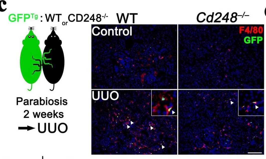
Cd248 disruption reduced infiltration and pro-fibrotic phenotype switching of kidney macrophages during renal fibrosis. (a) Representative images of F4/80 immunostaining for macrophages in control and D14 UUO kidneys. F4/80+ staining was brown. Original magnification, × 100. Scale bar, 100 μm. (b) Quantification of F4/80+ areas on LPF images of kidney sections taken at 100 × magnification n = 10. (c). Representative images of green fluorescent protein (GFP)+ circulation-derived cells and F4/80+ (red) macrophages in control and day 3 (D3) UUO-kidneys of WT and Cd248–/– parabionts joined surgically to transgenic GFP (GFPTg) mice. Arrowhead indicates an F4/80+GFP+ macrophage. Original magnification, × 100. Scale bar, 100 μm. (d) Quantification of GFP+, F4/80+ and F4/80+GFP+ cells on LPF images of control (upper panel) and D3 UUO (lower panel) kidneys of WT and Cd248–/– parabionts. n = 3. (e) qPCR of genes encoding cytokines and enzymes in macrophages isolated from D7 UUO kidneys. n = 4. (f) qPCR of Nos2, Arg1 and Ccl17 in lipopolysaccharide (LPS) and interferon gamma (IFN gamma)–primed RAW264.7 macrophages co-cultured with D7 UUO-kidney myofibroblasts isolated from WT or Cd248–/– mice in the Transwell system. RAW264.7 macrophages co-cultured with medium were used as control. n = 4. (g) qPCR of Ccl17 in WT bone marrow–derived macrophages (BMDMs, Mϕ) co-cultured with medium only (control) or with WT or Cd248–/– UUO-kidney myofibroblasts (MF) in the same dish. Recombinant CD248 (rCD248) was included in the culture as indicated. n = 5. Data are expressed as means ± standard errors of the mean. *P < 0.05, **P < 0.01, ***P < 0.001 by one-way ANOVA with post hoc Tukey’s multiple comparisons test in (b,g) and unpaired t-test in (d,e,f). Image collected and cropped by CiteAb from the following open publication (https://pubmed.ncbi.nlm.nih.gov/33033277), licensed under a CC-BY license. Not internally tested by R&D Systems.Detection of Mouse CCL17/TARC by Immunocytochemistry/ Immunofluorescence
Cd248 disruption reduced infiltration and pro-fibrotic phenotype switching of kidney macrophages during renal fibrosis. (a) Representative images of F4/80 immunostaining for macrophages in control and D14 UUO kidneys. F4/80+ staining was brown. Original magnification, × 100. Scale bar, 100 μm. (b) Quantification of F4/80+ areas on LPF images of kidney sections taken at 100 × magnification n = 10. (c). Representative images of green fluorescent protein (GFP)+ circulation-derived cells and F4/80+ (red) macrophages in control and day 3 (D3) UUO-kidneys of WT and Cd248–/– parabionts joined surgically to transgenic GFP (GFPTg) mice. Arrowhead indicates an F4/80+GFP+ macrophage. Original magnification, × 100. Scale bar, 100 μm. (d) Quantification of GFP+, F4/80+ and F4/80+GFP+ cells on LPF images of control (upper panel) and D3 UUO (lower panel) kidneys of WT and Cd248–/– parabionts. n = 3. (e) qPCR of genes encoding cytokines and enzymes in macrophages isolated from D7 UUO kidneys. n = 4. (f) qPCR of Nos2, Arg1 and Ccl17 in lipopolysaccharide (LPS) and interferon gamma (IFN gamma)–primed RAW264.7 macrophages co-cultured with D7 UUO-kidney myofibroblasts isolated from WT or Cd248–/– mice in the Transwell system. RAW264.7 macrophages co-cultured with medium were used as control. n = 4. (g) qPCR of Ccl17 in WT bone marrow–derived macrophages (BMDMs, Mϕ) co-cultured with medium only (control) or with WT or Cd248–/– UUO-kidney myofibroblasts (MF) in the same dish. Recombinant CD248 (rCD248) was included in the culture as indicated. n = 5. Data are expressed as means ± standard errors of the mean. *P < 0.05, **P < 0.01, ***P < 0.001 by one-way ANOVA with post hoc Tukey’s multiple comparisons test in (b,g) and unpaired t-test in (d,e,f). Image collected and cropped by CiteAb from the following open publication (https://pubmed.ncbi.nlm.nih.gov/33033277), licensed under a CC-BY license. Not internally tested by R&D Systems.Applications for Mouse CCL17/TARC Antibody
Western Blot
Sample: Recombinant Mouse CCL17/TARC (Catalog # 529-TR)
Neutralization
Reviewed Applications
Read 2 reviews rated 5 using AF529 in the following applications:
Formulation, Preparation, and Storage
Purification
Reconstitution
Formulation
Shipping
Stability & Storage
- 12 months from date of receipt, -20 to -70 °C as supplied.
- 1 month, 2 to 8 °C under sterile conditions after reconstitution.
- 6 months, -20 to -70 °C under sterile conditions after reconstitution.
Background: CCL17/TARC
Human thymus and activation-regulated chemokine (TARC) also known as CCL17, is a novel CC chemokine identified using a signal sequence trap method. Mouse TARC was discovered as a dendritic cell (DC) specific gene by differentiation RNA display. Mouse TARC cDNA encodes a highly basic 93 amino acid (aa) residue precursor protein with a 23 aa residue putative signal peptide that is cleaved to generate the 70 aa residue mature secreted protein. Among CC chemokine family members, TARC has approximately 24 - 29% amino acid sequence identity with RANTES, MIP-1 alpha, MIP-1 beta, MCP-1, MCP-2, MCP-3 and I-309. The gene for human TARC has been mapped to chromosome 16q13 rather than chromosome 17 where the genes for many human CC chemokines are clustered. Mouse TARC is constitutively expressed in thymic DC, and at a lower level in lymph node DC in the lung. Recombinant TARC has been shown to be chemotactic for T cell lines and antigen-primed T helper cells. In humans, TARC was identified to be a specific functional ligand for CCR-4 and CCR-8, receptors that are selectively expressed on T cells.
References
- Imai, T. et al. (1997) J. Biol. Chem. 272:15036.
- Imai, T. et al. (1996) J. Biol. Chem. 271:21514.
- Nomiyama, H. et al. (1997) Genomics 40:211.
- Lieberam, I. et al. (1999) Eur. J. Immunol. 29:2684.
Alternate Names
Gene Symbol
UniProt
Additional CCL17/TARC Products
Product Documents for Mouse CCL17/TARC Antibody
Product Specific Notices for Mouse CCL17/TARC Antibody
For research use only

