Mouse CCL4/MIP-1 beta Antibody
R&D Systems, part of Bio-Techne | Catalog # AF-451-NA


Key Product Details
Validated by
Biological Validation
Species Reactivity
Validated:
Mouse
Cited:
Mouse, Rat, Transgenic Mouse
Applications
Validated:
Immunocytochemistry, Immunohistochemistry, Neutralization, Western Blot
Cited:
ELISA Development, Flow Cytometry, Immunocytochemistry, Immunohistochemistry, Immunohistochemistry-Paraffin, Neutralization
Label
Unconjugated
Antibody Source
Polyclonal Goat IgG
Product Specifications
Immunogen
E. coli-derived recombinant mouse CCL4/MIP-1 beta
Ala24-Asn92
Accession # Q5QNV9
Ala24-Asn92
Accession # Q5QNV9
Specificity
Detects CCL4/MIP-1 beta in direct ELISAs and Western blots. In direct ELISAs, less than 5% cross-reactivity with recombinant human (rh) MIP-1 beta and recombinant mouse (rm) MIP-1 alpha is observed. This antibody will not neutralize the biological activity of rmMIP-1 alpha. In the chemotaxis assay this antibody will partially neutralize rhMIP-1 alpha and rhMIP-1 beta at a 10‑30 fold higher IgG concentration.
Clonality
Polyclonal
Host
Goat
Isotype
IgG
Endotoxin Level
<0.10 EU per 1 μg of the antibody by the LAL method.
Scientific Data Images for Mouse CCL4/MIP-1 beta Antibody
Detection of Mouse CCL4/MIP‑1 beta by Western Blot.
Western blot shows lysates of RAW 264.7 mouse monocyte/macrophage cell line untreated (-) or treated (+) with 10 µg/mL LPS for 4 hours. PVDF membrane was probed with 1 µg/mL of Goat Anti-Mouse CCL4/MIP-1 beta Antigen Affinity-purified Polyclonal Antibody (Catalog # AF-451-NA) followed by HRP-conjugated Anti-Goat IgG Secondary Antibody (Catalog # HAF017). A specific band was detected for CCL4/MIP-1 beta at approximately 12 kDa (as indicated). This experiment was conducted under reducing conditions and using Immunoblot Buffer Group 1.CCL4/MIP‑1 beta in RAW 264.7 Mouse Cell Line.
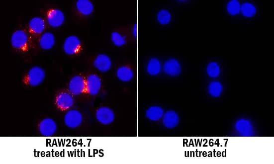
CCL4/MIP-1 beta was detected in immersion fixed RAW 264.7 mouse monocyte/macrophage cell line untreated (right panel) and treated with LPS (left panel) using Goat Anti-Mouse CCL4/MIP-1 beta Antigen Affinity-purified Polyclonal Antibody (Catalog # AF-451-NA) at 1.7 µg/mL for 3 hours at room temperature. Cells were stained using the NorthernLights™ 557-conjugated Anti-Goat IgG Secondary Antibody (red; Catalog # NL001) and counterstained with DAPI (blue). Specific staining was localized to cytoplasm. View our protocol for Fluorescent ICC Staining of Cells on Coverslips.CCL4/MIP‑1 beta in Mouse Small Intestine.
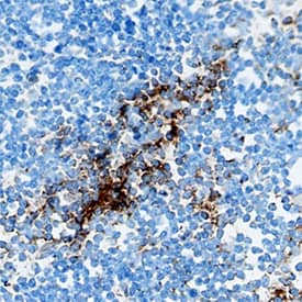
CCL4/MIP-1 beta was detected in perfusion fixed frozen sections of mouse small intestine using Goat Anti-Mouse CCL4/MIP-1 beta Antigen Affinity-purified Polyclonal Antibody (Catalog # AF-451-NA) at 1.7 µg/mL overnight at 4 °C. Tissue was stained using the Anti-Goat HRP-DAB Cell & Tissue Staining Kit (brown; Catalog # CTS008) and counterstained with hematoxylin (blue). Specific staining was localized to Peyer's patches. View our protocol for Chromogenic IHC Staining of Frozen Tissue Sections.Applications for Mouse CCL4/MIP-1 beta Antibody
Application
Recommended Usage
Immunocytochemistry
1-15 µg/mL
Sample: Immersion fixed RAW 264.7 mouse monocyte/macrophage cell line treated with LPS
Sample: Immersion fixed RAW 264.7 mouse monocyte/macrophage cell line treated with LPS
Immunohistochemistry
1-15 µg/mL
Sample: Perfusion fixed frozen sections of mouse thymus and mouse small intestine
Sample: Perfusion fixed frozen sections of mouse thymus and mouse small intestine
Western Blot
1 µg/mL
Sample: RAW 264.7 mouse monocyte/macrophage cell line treated with LPS
Sample: RAW 264.7 mouse monocyte/macrophage cell line treated with LPS
Neutralization
Measured by its ability to neutralize CCL4/MIP-1 beta-induced chemotaxis in the BaF3 mouse pro-B cell line transfected with human CCR5. The Neutralization Dose (ND50) is typically 0.2-1.0 µg/mL in the presence of 25 ng/mL Recombinant Mouse CCL4/MIP-1 beta.
Formulation, Preparation, and Storage
Purification
Antigen Affinity-purified
Reconstitution
Reconstitute at 0.2 mg/mL in sterile PBS. For liquid material, refer to CoA for concentration.
Formulation
Lyophilized from a 0.2 μm filtered solution in PBS with Trehalose. *Small pack size (SP) is supplied either lyophilized or as a 0.2 µm filtered solution in PBS.
Shipping
Lyophilized product is shipped at ambient temperature. Liquid small pack size (-SP) is shipped with polar packs. Upon receipt, store immediately at the temperature recommended below.
Stability & Storage
Use a manual defrost freezer and avoid repeated freeze-thaw cycles.
- 12 months from date of receipt, -20 to -70 °C as supplied.
- 1 month, 2 to 8 °C under sterile conditions after reconstitution.
- 6 months, -20 to -70 °C under sterile conditions after reconstitution.
Background: CCL4/MIP-1 beta
References
- Rot, A. and U.H. von Andrian (2004) Annu. Rev. Immunol. 22:891.
- Menten, P. et al. (2002) Cytokine Growth Factor Rev. 13:455.
- Sun, X. et al. (2006) Infec. Immun. 74:5943.
- Bisset, L.R. and Schmid-Grendelmeier, P. (2005) Curr. Opin. Pulm. Med. 11:35.
- Frangogiannis, N.G. (2004) Inflamm. Res. 53:585.
- Bystry, R.S. et al. (2001) Nat. Immunol. 2:1126.
- Oliveira, S.H.P. et al. (2002) J. Leukoc. Biol. 71:1019.
- Schall, T.J. et al. (1993) J. Exp. Med. 177:1821.
- Guan, E. et al. (2001) J. Biol. Chem. 276:12404.
- Guan, E. et al. (2002) J. Biol. Chem. 277:32348.
- Guan, E. et al. (2004) J. Cell. Biochem. 92:53.
Alternate Names
Exodus-3, MIP-1 beta, MIP1 beta
Entrez Gene IDs
Gene Symbol
CCL4
UniProt
Additional CCL4/MIP-1 beta Products
Product Documents for Mouse CCL4/MIP-1 beta Antibody
Product Specific Notices for Mouse CCL4/MIP-1 beta Antibody
For research use only
Loading...
Loading...
Loading...
Loading...


