Mouse CCR7 APC-conjugated Antibody
R&D Systems, part of Bio-Techne | Catalog # FAB3477A


Conjugate
Catalog #
Key Product Details
Validated by
Biological Validation
Species Reactivity
Mouse
Applications
Flow Cytometry
Label
Allophycocyanin (Excitation = 620-650 nm, Emission = 660-670 nm)
Antibody Source
Monoclonal Rat IgG2A Clone # 4B12
Product Specifications
Immunogen
RBC-2H3 cells expressing mouse CCR7
Accession # NP_031745
Accession # NP_031745
Specificity
Detects mouse CCR7.
Clonality
Monoclonal
Host
Rat
Isotype
IgG2A
Scientific Data Images for Mouse CCR7 APC-conjugated Antibody
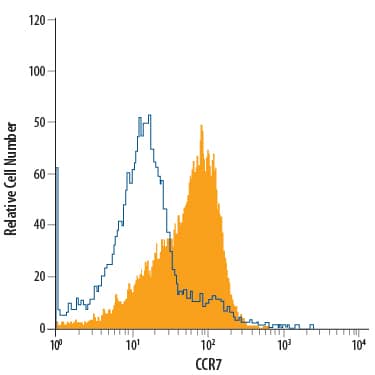
Detection of CCR7 in Mouse CD4+Splenocytes by Flow Cytometry.
Mouse CD4+splenocytes were stained with Rat Anti-Mouse CCR7 APC-conjugated Monoclonal Antibody (Catalog # FAB3477A, filled histogram) or isotype control antibody (Catalog # IC006A, open histogram). View our protocol for Staining Membrane-associated Proteins.Detection of Mouse CCR7 by Flow Cytometry
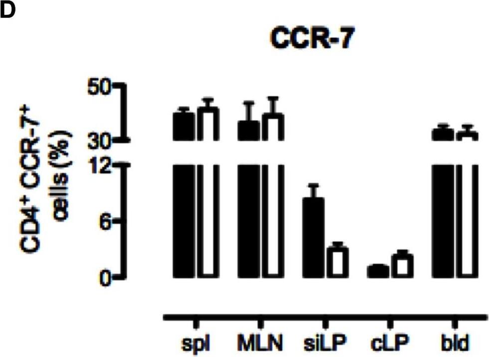
B6 and CD69−/− CD4 T cells differ in the expression of naïve and memory surface cell markers.Cells were isolated from the spleen (spl), mesenteric lymph nodes (MLN), small intestinal lamina propria (siLP), colonic lamina propria (cLP) and blood (bld) of non-treated B6 and CD69−/− mice and analyzed by flow cytometry. The surface expression of CD44 (A), CD103 (B), CD62L (C), CCR-7 (D), CCR-9 (E) and alpha4 beta7 integrin (F) by CD4 T cells were analysed. Graphs represent mean (± SEM) of CD4 T cell fraction expressing the indicated molecule for four mice per each strain per tissue. Black bars represent data for B6 and white bars for CD69−/− cells. *p≤0.05. Image collected and cropped by CiteAb from the following publication (https://pubmed.ncbi.nlm.nih.gov/23776480), licensed under a CC-BY license. Not internally tested by R&D Systems.Applications for Mouse CCR7 APC-conjugated Antibody
Application
Recommended Usage
Flow Cytometry
10 µL/106 cells
Sample: Mouse CD4+ splenocytes
Sample: Mouse CD4+ splenocytes
Formulation, Preparation, and Storage
Purification
Protein A or G purified from hybridoma culture supernatant
Formulation
Supplied in a saline solution containing BSA and Sodium Azide.
Shipping
The product is shipped with polar packs. Upon receipt, store it immediately at the temperature recommended below.
Stability & Storage
Protect from light. Do not freeze.
- 12 months from date of receipt, 2 to 8 °C as supplied.
Background: CCR7
Alternate Names
BLR2, CC-CKR-7, CCR7, CD197, CDw197, CMKBR7, EBI1
Gene Symbol
CCR7
UniProt
Additional CCR7 Products
Product Documents for Mouse CCR7 APC-conjugated Antibody
Product Specific Notices for Mouse CCR7 APC-conjugated Antibody
For research use only
Loading...
Loading...
Loading...
Loading...
Loading...
