Mouse CD1d1 Antibody
R&D Systems, part of Bio-Techne | Catalog # AF4884

Key Product Details
Species Reactivity
Applications
Label
Antibody Source
Product Specifications
Immunogen
Gln22-Gly305
Accession # NP_031665
Specificity
Clonality
Host
Isotype
Scientific Data Images for Mouse CD1d1 Antibody
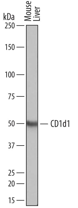
Detection of Mouse CD1d1 by Western Blot.
Western blot shows lysates of mouse liver tissue. PVDF membrane was probed with 0.5 µg/mL of Sheep Anti-Mouse CD1d1 Antigen Affinity-purified Polyclonal Antibody (Catalog # AF4884) followed by HRP-conjugated Anti-Sheep IgG Secondary Antibody (Catalog # HAF016). A specific band was detected for CD1d1 at approximately 50 kDa (as indicated). This experiment was conducted under reducing conditions and using Immunoblot Buffer Group 1.Applications for Mouse CD1d1 Antibody
Western Blot
Sample: Mouse liver tissue
Formulation, Preparation, and Storage
Purification
Reconstitution
Formulation
Shipping
Stability & Storage
- 12 months from date of receipt, -20 to -70 °C as supplied.
- 1 month, 2 to 8 °C under sterile conditions after reconstitution.
- 6 months, -20 to -70 °C under sterile conditions after reconstitution.
Background: CD1d1
CD1d is a 48 kDa transmembrane glycoprotein that belongs to the CD1 family of glycolipid antigen-presenting MHC-like molecules. In mouse, there are two closely related CD1d genes, CD1d1 and CD1d2, whereas human and rat have only one (1, 2). Mature mouse CD1d1 consists of a 284 amino acid (aa) extracellular domain (ECD) with one Ig-like domain, a 21 aa transmembrane segment, and a 10 aa cytoplasmic tail (3). Within the ECD, mouse CD1d1 shares 94% aa sequence identity with mouse CD1d2, and 65% and 87% with human and rat CD1d, respectively. Complexes of CD1d1 with beta2-microglobulin and endogenous glycolipids are constitutively expressed on antigen presenting cells, cortical thymocytes, liver sinusoidal endothelial cells, Kupffer cells, and hepatocytes (1). A cytoplasmic motif mediates CD1d1 recycling through the endosomal/lysosomal system where it is loaded with processed exogenous glycolipids by saposin lipid transfer proteins (4 - 8). CD1d1-presented glycolipids are recognized by canonical NKT cells that utilize an invariant V alpha14-J alpha18 chain in their T cell receptor (V alpha24-J alpha18 in human) (9, 10). NKT cells that express V chains other than alpha14 can also recognize CD1d1-presented glycolipids but do not require them to be endosomally loaded (10, 11). NKT cells respond to a variety of CD1d1-presented glycolipids including alpha-galactosylceramide ( alpha-GalCer), a structural relative of microbial cell wall components, and the endogenous isoglobotrihexosylceramide (iGb3) (2, 9, 12). The interaction with glycolipid-loaded CD1d1 is critical for NKT cell development and induces their rapid secretion of both Th1 and Th2 type cytokines (10, 11, 13, 14). In humans, infection with HSV-1 suppresses NKT cell activation by blocking the intracellular cycling of CD1d in antigen presenting cells (15).
References
- Bendelac, A. et al. (2007) Annu. Rev. Immunol. 25:297.
- Brutkiewicz, R.R. (2006) J. Immunol. 177:769.
- Balk, S.P. et al. (1991) J. Immunol. 146:768.
- De Silva, A.D. et al. (2002) J. Immunol. 168:723.
- Roberts, T.J. et al. (2002) J. Immunol. 168:5409.
- Kang, S.J. and P. Cresswell (2004) Nat. Immunol. 5:175.
- Zhou, D. et al. (2004) Science 303:523.
- Prigozy, T.O. et al. (2001) Science 291:664.
- Kawano, T. et al. (1997) Science 278:1626.
- Chiu, Y.H. et al. (1999) J. Exp. Med. 189:103.
- Behar, S.M. et al. (1999) J. Immunol. 162:161.
- Zhou, D. et al. (2004) Science 306:1786.
- Mendiratta, S.K. et al. (1997) Immunity 6:469.
- Stanic, A.K. et al. (2003) J. Immunol. 171:4539.
- Yuan, W. et al. (2006) Nat. Immunol. 7:835.
Alternate Names
Gene Symbol
UniProt
Additional CD1d1 Products
Product Documents for Mouse CD1d1 Antibody
Product Specific Notices for Mouse CD1d1 Antibody
For research use only
