Mouse CD28 Antibody
R&D Systems, part of Bio-Techne | Catalog # MAB4832


Key Product Details
Species Reactivity
Validated:
Cited:
Applications
Validated:
Cited:
Label
Antibody Source
Product Specifications
Immunogen
Accession # P31041
Specificity
Clonality
Host
Isotype
Endotoxin Level
Scientific Data Images for Mouse CD28 Antibody
Mouse CD28 Antibody Induces Proliferation in Mouse T Cells.
Rat Anti-Mouse CD28 Monoclonal Antibody (Catalog # MAB4832) induces proliferation in mouse T cells in the presence of 100 ng/mL Hamster Anti-Mouse CD3e Monoclonal Antibody (Catalog # MAB484), in a dose dependent manner, as measured by Resazurin (Catalog # AR002), The ED50 for this effect is typically 0.07-0.42 µg/mL.Detection of CD28 in Mouse Thymocytes by Flow Cytometry.
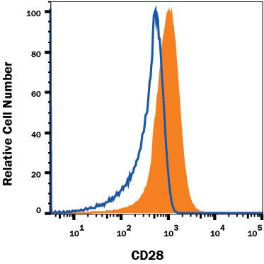
Mouse thymocytes were stained with Rat Anti-Mouse CD28 Monoclonal Antibody (Catalog # MAB4832, filled histogram) or isotype control antibody (MAB005, open histogram), followed by Phycoerythrin-conjugated Anti-Rat IgG Secondary Antibody (F0105B). Staining was performed using our Membrane-Associated Proteins protocol.Detection of CD28 in CD3+Mouse Splenocytes by Flow Cytometry.
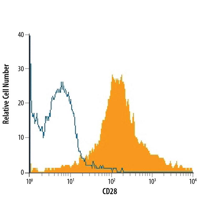
CD3+mouse splenocytes were stained with Rat Anti-Mouse CD28 Monoclonal Antibody (Catalog # MAB4832, filled histogram) or isotype control antibody (Catalog # MAB005, open histogram), followed by Phycoerythrin-conjugated Anti-Rat IgG Secondary Antibody (Catalog # F0105B).Applications for Mouse CD28 Antibody
Agonist Activity
Sample: Mouse T cells
CyTOF-ready
Flow Cytometry
Sample: CD3+ mouse splenocytes and mouse thymocytes
Formulation, Preparation, and Storage
Purification
Reconstitution
Formulation
Shipping
Stability & Storage
- 12 months from date of receipt, -20 to -70 °C as supplied.
- 1 month, 2 to 8 °C under sterile conditions after reconstitution.
- 6 months, -20 to -70 °C under sterile conditions after reconstitution.
Background: CD28
CD28 and CTLA-4, together with their ligands B7-1 and B7-2, constitute one of the dominant costimulatory pathways that regulate T and B cell responses. CD28 and CTLA-4 are structurally homologous molecules that are members of the immunoglobulin (Ig) gene superfamily. Both CD28 and CTLA-4 are composed of a single Ig
V‑like extracellular domain, a transmembrane domain and an intracellular domain. CD28 and CTLA-4 are both expressed on the cell surface as disulfide-linked homodimers or as monomers. The genes encoding these two molecules are closely linked on human chromosome 2 and mouse chromosome 1. Mouse CD28 is expressed constitutively on virtually 100% of mouse T cells and on developing thymocytes. Cell surface expression of mouse CD28 is down-regulated upon ligation of CD28 in the presence of PMA or PHA. In contrast, CTLA-4 is not expressed constitutively but is upregulated rapidly following T cell activation and CD28 ligation. Cell surface expression of CTLA-4 peaks approximately 48 hours after activation. Although both CTLA-4 and CD28 can bind to the same ligands, CTLA-4 binds to
B7‑1 and B7‑2 with a 20-100 fold higher affinity than CD28. CD28/B7 interaction has been shown to prevent apoptosis of activated T cells via the up-regulation of
Bcl‑xL. CD28 ligation has also been shown to regulate Th1/Th2 differentiation. Agonist activity has been reported using MAB4831 (4,5).
References
- Lenschow, D.J. et al. (1996) Annu. Rev. Immunol. 14:233.
- Hathcock, K.S. and R.J. Hodes (1996) Advances in Immunol. 62:131.
- Ward, S.G. (1996) Biochem. J. 318:361.
- Nguyen, P. et al. (2003) Blood 13:4320.
- Orbach, A. et al. (2007) J. Immunol. 179:7287.
Alternate Names
Gene Symbol
UniProt
Additional CD28 Products
Product Documents for Mouse CD28 Antibody
Product Specific Notices for Mouse CD28 Antibody
For research use only

