Mouse CD36/SR-B3 Antibody
R&D Systems, part of Bio-Techne | Catalog # MAB25191


Key Product Details
Species Reactivity
Validated:
Cited:
Applications
Validated:
Cited:
Label
Antibody Source
Product Specifications
Immunogen
Gly30-Lys439
Accession # Q08857
Specificity
Clonality
Host
Isotype
Scientific Data Images for Mouse CD36/SR-B3 Antibody
Detection of CD36/SR‑B3 in J774A.1 Mouse Cell Line by Flow Cytometry.
J774A.1 mouse reticulum cell sarcoma macrophage cell line was stained with Rat Anti-Mouse CD36/SR-B3 Monoclonal Antibody (Catalog # MAB25191, filled histogram) or isotype control antibody (Catalog # MAB005, open histogram), followed by Allophycocyanin-conjugated Anti-Rat IgG Secondary Antibody (Catalog # F0113).CD36/SR‑B3 in Mouse Heart.
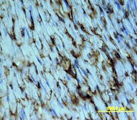
CD36/SR-B3 was detected in immersion fixed frozen sections of mouse heart using Rat Anti-Mouse CD36/SR-B3 Monoclonal Antibody (Catalog # MAB25191) at 15 µg/mL overnight at 4 °C. Tissue was stained using the Anti-Rat HRP-DAB Cell & Tissue Staining Kit (brown; Catalog # CTS017) and counterstained with hematoxylin (blue). Specific staining was localized to cell surfaces and cytoplasm. View our protocol for Chromogenic IHC Staining of Frozen Tissue Sections.CD36/SR‑B3 in Mouse Thymus.
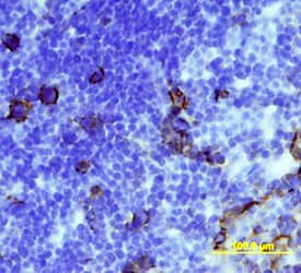
CD36/SR-B3 was detected in immersion fixed frozen sections of mouse thymus using Rat Anti-Mouse CD36/SR-B3 Monoclonal Antibody (Catalog # MAB25191) at 15 µg/mL overnight at 4 °C. Tissue was stained using the Anti-Rat HRP-DAB Cell & Tissue Staining Kit (brown; Catalog # CTS017) and counterstained with hematoxylin (blue). Specific staining was localized to cell surfaces and cytoplasm. View our protocol for Chromogenic IHC Staining of Frozen Tissue Sections.Applications for Mouse CD36/SR-B3 Antibody
CyTOF-ready
Flow Cytometry
Sample: J774A.1 mouse reticulum cell sarcoma macrophage cell line
Immunohistochemistry
Sample: Immersion fixed frozen sections of mouse heart and thymus
Formulation, Preparation, and Storage
Purification
Reconstitution
Formulation
Shipping
Stability & Storage
- 12 months from date of receipt, -20 to -70 °C as supplied.
- 1 month, 2 to 8 °C under sterile conditions after reconstitution.
- 6 months, -20 to -70 °C under sterile conditions after reconstitution.
Background: CD36/SR-B3
CD36 (alternatively known as platelet membrane glycoprotein IV (GPIV), thrombospondin receptor, fatty acid translocase (FAT), and scavenger receptor class B, member 3 (SR-B3)) is an 88 kDa, integral membrane glycoprotein that belongs to the class B scavenger receptor family (1, 2). The molecule is described as being ditopic, with two transmembrane segments connected by an extracellular loop (3). Mouse CD36 is synthesized as a 472 amino acid (aa) protein that contains a 6 aa N‑terminal cytoplasmic domain, a 22 aa N‑terminal transmembrane segment, a 420 aa extracellular “loop”, a 22 aa C‑terminal transmembrane segment, and a 9 aa C‑terminal cytoplasmic tail (4). Both cytoplasmic tails are palmitoylated, with the C‑terminal tail involved in oxidized LDL binding (5, 6). With respect to the extracellular loop, the N‑terminal region is believed to bind both thrombospondin-1 and Plasmodium-infected erythrocytes. Other ligands for CD36 include long-chain fatty acids, collagen, phospholipids and apoptotic cells (1). The extracellular loop of mouse CD36 shares 94%, 92%, 84% and 84% aa sequence identity with the extracellular loops of rat, hamster, human and bovine CD36, respectively. Cells known to express CD36 include capillary endothelium, adipocytes, skeletal muscle cells, intestinal epithelium, smooth muscle cells and hematopoietic cells such as RBC’s, platelets and monocytes (1). On the surface of cells, CD36 is suggested to exist as a dimer in response to ligation (7). CD36 is reported to regulate fatty uptake, act as an angiogenic with TSP-1, and participate in the clearance of apoptotic phagocytes (1, 8).
References
- Febbraio, M. et al. (2001) J. Clin. Invest. 108:795.
- Silverstein, R.L. and M. Febbraio (2000) Curr. Opin. Lipid. 11:483.
- Gruarin, P. et al. (2000) Biochem. Biophys. Res. Commun. 275:446.
- Endemann, G. et al. (1993) J. Biol. Chem. 268:11811.
- Malaud, E. et al. (2002) Biochem. J. 364:507.
- Tao, N. et al. (1996) J. Biol. Chem. 271:22315.
- Daviet, L. et al. (1997) Thromb. Haemost. 78:897.
- Simantov, R. and R.L. Silverstein (2003) Front. Biosci. 8:s874.
Long Name
Alternate Names
Gene Symbol
UniProt
Additional CD36/SR-B3 Products
Product Documents for Mouse CD36/SR-B3 Antibody
Product Specific Notices for Mouse CD36/SR-B3 Antibody
For research use only

