Mouse CD55/DAF Antibody
R&D Systems, part of Bio-Techne | Catalog # MAB5376


Key Product Details
Species Reactivity
Validated:
Cited:
Applications
Validated:
Cited:
Label
Antibody Source
Product Specifications
Immunogen
Met1-Pro359
Accession # Q61475
Specificity
Clonality
Host
Isotype
Scientific Data Images for Mouse CD55/DAF Antibody
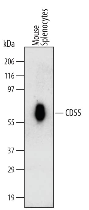
Detection of Mouse CD55/DAF by Western Blot.
Western blot shows lysates of mouse splenocytes. PVDF Membrane was probed with 2 µg/mL of Rat Anti-Mouse CD55/DAF Monoclonal Antibody (Catalog # MAB5376) followed by HRP-conjugated Anti-Rat IgG Secondary Antibody (Catalog # HAF005). A specific band was detected for CD55/DAF at approximately 60 kDa (as indicated). This experiment was conducted under non-reducing conditions and using Immunoblot Buffer Group 1.Detection of Mouse CD55/DAF by Western Blot.
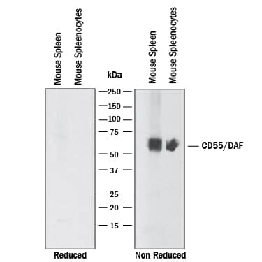
Western blot shows lysates of mouse spleen tissue and mouse splenocytes. PVDF membrane was probed with 2 µg/mL of Rat Anti-Mouse CD55/DAF Monoclonal Antibody (Catalog # MAB5376) followed by HRP-conjugated Anti-Rat IgG Secondary Antibody (Catalog # HAF005). A specific band was detected for CD55/DAF at approximately 75 kDa (as indicated). This experiment was conducted under non-reducing conditions and using Immunoblot Buffer Group 1. No bands were observed when using reducing conditions.Detection of CD55/DAF in Mouse Splenocytes by Flow Cytometry.
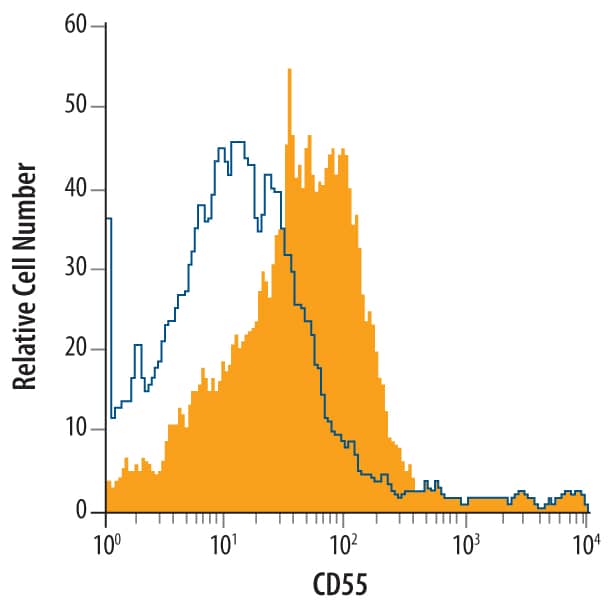
Mouse splenocytes were stained with Rat Anti-Mouse CD55/DAF Monoclonal Antibody (Catalog # MAB5376, filled histogram) or isotype control antibody (Catalog # MAB006, open histogram), followed by Phycoerythrin-conjugated Anti-Rat IgG F(ab')2Secondary Antibody (Catalog # F0105B).Applications for Mouse CD55/DAF Antibody
CyTOF-ready
Flow Cytometry
Sample: Mouse splenocytes
Western Blot
Sample: Mouse spleen tissue and Mouse splenocytes
under non-reducing conditions only
Formulation, Preparation, and Storage
Purification
Reconstitution
Formulation
Shipping
Stability & Storage
- 12 months from date of receipt, -20 to -70 °C as supplied.
- 1 month, 2 to 8 °C under sterile conditions after reconstitution.
- 6 months, -20 to -70 °C under sterile conditions after reconstitution.
Background: CD55/DAF
CD55 (Decay-accelarating factor/DAF) is a glycoprotein member of the RCA family of molecules. It is found on blood cells, epithelium and endothelium, and serves both as a receptor for CD97, and a negative regulator of the C3 convertases, C4b2a and C3bBb. Mature mouse CD55 is the product of two genes that arose by duplication. There is a 55-60 kDa, 356 amino acid (aa), GPI-linked form that is ubiquitously expressed. This molecule contains four SUSHI domains (aa 35-285), a Ser/Thr-rich region (aa 288-362), and a GPI-anchor at Gly362. There is also a 50 kDa, 379 aa, type I transmembrane form that is testis-associated. It shows the same domain architecture and is 93% aa identical to the GPI-form. At least four GPI gene isoforms exist. They diverge after Ile285 and show deletions and substitutions. Over aa 35-359, mouse CD55 is 66% and 50% aa identical to rat and human CD55, respectively.
Alternate Names
Gene Symbol
UniProt
Additional CD55/DAF Products
Product Documents for Mouse CD55/DAF Antibody
Product Specific Notices for Mouse CD55/DAF Antibody
For research use only

