Mouse CD83 Antibody
R&D Systems, part of Bio-Techne | Catalog # AF1437

Key Product Details
Species Reactivity
Validated:
Cited:
Applications
Validated:
Cited:
Label
Antibody Source
Product Specifications
Immunogen
Met22-Ala134
Accession # O88324
Specificity
Clonality
Host
Isotype
Endotoxin Level
Scientific Data Images for Mouse CD83 Antibody
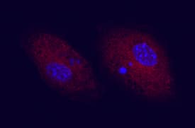
CD83 in Mouse Splenocytes.
CD83 was detected in immersion fixed mouse splenocytes using 10 µg/mL Goat Anti-Mouse CD83 Antigen Affinity-purified Polyclonal Antibody (Catalog # AF1437) for 3 hours at room temperature. Cells were stained with the NorthernLights™ 557-conjugated Anti-Goat IgG Secondary Antibody (red; Catalog # NL001) and counterstained with DAPI (blue). View our protocol for Fluorescent ICC Staining of Non-adherent Cells.CD83 in Mouse Dendritic Cells.
CD83 was detected in immersion fixed LPS-stimulated mouse dendritic cells using Goat Anti-Mouse CD83 Antigen Affinity-purified Polyclonal Antibody (Catalog # AF1437) at 10 µg/mL for 3 hours at room temperature. Cells were stained using the NorthernLights™ 557-conjugated Anti-Goat IgG Secondary Antibody (red; Catalog # NL001) and counterstained with DAPI (blue). View our protocol for Fluorescent ICC Staining of Non-adherent Cells.Applications for Mouse CD83 Antibody
Adhesion Blockade
CyTOF-ready
Flow Cytometry
Sample: LPS-treated mature mouse dendritic cells
Immunocytochemistry
Sample: Immersion fixed mouse splenocytes and dendritic cells
Western Blot
Sample: Recombinant Mouse CD83 Fc Chimera (Catalog # 1437-CD)
Reviewed Applications
Read 1 review rated 5 using AF1437 in the following applications:
Formulation, Preparation, and Storage
Purification
Reconstitution
Formulation
Shipping
Stability & Storage
- 12 months from date of receipt, -20 to -70 °C as supplied.
- 1 month, 2 to 8 °C under sterile conditions after reconstitution.
- 6 months, -20 to -70 °C under sterile conditions after reconstitution.
Background: CD83
Mouse CD83 is a 30‑35 kDa member of the Siglec (or sialic-acid-binding immunoglobulin-like lectin) family of transmembrane proteins (1, 2, 3). CD83 is synthesized as a type I transmembrane glycoprotein that contains a 114 amino acid (aa) extracellular region, a 22 aa transmembrane segment, and a 39 aa cytoplasmic domain. It contains one V type Ig-like domain in the extracellular region with no inhibitory cytoplasmic motif(s). In the extracellular region, mouse and human CD83 are 66% aa identical (1, 2, 4). Relative to mouse, human CD83 has an 11 aa insertion in its extracellular domain and is expressed as a 45‑55 kDa protein (1, 4, 5, 6). No alternate splice variants have been reported for mouse. In human, however, one soluble splice form has been reported and proteolytic processing is suggested to generate a second circulating isoform (6, 7). Notably, although soluble CD83 has the potential to exist as either a monomer or disulfide-linked dimer, both show immunosuppressive activity (4, 8, 9). Membrane CD83, by contrast, is immunostimulatory (10). CD83 is a primary marker for dendritic cells (3, 5, 6). It is also found on B cells (6, 11), neutrophils (12), monocytes and macrophages (13). Except for dendritic cells, CD83 expression is often transient. CD83 binds to sialic acids on monocytes (3). The function of CD83 is only now becoming clear. As noted, membrane-immobilized CD83 appears to promote T cell proliferation, particularly of CD8+ cytotoxic T cells (14). On monocytes, CD83 may also drive monocytes into a fibrocyte phenotype (14). And a lack of membrane-expressed CD83 leads to an unusual IL-4/IL-10 producing CD4+ T cell phenotype (15).
References
- Berchtold, S. et. al. (1999) FEBS Lett. 461:211.
- Fujimoto, Y. and T.F. Tedder (2006) J. Med. Dent. Sci. 53:85.
- Scholler, N. et. al. (2001) J. Immunol. 166:3865.
- Lechmann, M. et al. (2005) Biochem. Biophys. Res. Commun. 329:132.
- Zhou, L-J. et. al. (1992) J. Immunol. 149:735.
- Hock, B.D. et al. (2001) Int. Immunol. 13:959.
- Dudziak, D. et al. (2005) J. Immunol. 174:6672.
- Kotzor, N. et al. (2004) Immunobiology 209:129.
- Zinser, E. et al. (2006) Immunobiology 21:449.
- Hirano, N. et al. (2006) Blood 107:1528.
- Cramer, S.O. et al. (2000) Int. Immunol. 12:1347.
- Yamashiro, S. et al. (2000) Blood 96:3958.
- Cao, W. et al. (2005) Biochem. J. 385:85.
- Scholler, N. et al. (2002) J. Immunol. 168:2599.
- Garcia-Martinez, L.F. et al. (2004) J. Immunol. 173:2995.
Alternate Names
Gene Symbol
UniProt
Additional CD83 Products
Product Documents for Mouse CD83 Antibody
Product Specific Notices for Mouse CD83 Antibody
For research use only

