Mouse CHL-1/L1CAM-2 Antibody
R&D Systems, part of Bio-Techne | Catalog # AF2147


Key Product Details
Validated by
Species Reactivity
Validated:
Cited:
Applications
Validated:
Cited:
Label
Antibody Source
Product Specifications
Immunogen
Ala25-Gln1043 (Leu227-Gln242 del, Ala243Ser)
Accession # BAC30699
Specificity
Clonality
Host
Isotype
Scientific Data Images for Mouse CHL-1/L1CAM-2 Antibody
Detection of Mouse CHL‑1/L1CAM‑2 by Western Blot.
Western blot shows lysates of mouse brain stem tissue. PVDF membrane was probed with 0.25 µg/mL of Goat Anti-Mouse CHL-1/L1CAM-2 Antigen Affinity-purified Polyclonal Antibody (Catalog # AF2147) followed by HRP-conjugated Anti-Goat IgG Secondary Antibody (Catalog # HAF019). A specific band was detected for CHL-1/L1CAM-2 at approximately 200 kDa (as indicated). This experiment was conducted under reducing conditions and using Immunoblot Buffer Group 1.Detection of Mouse CHL‑1/L1CAM‑2 by Simple WesternTM.
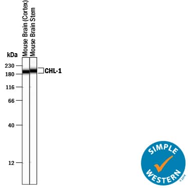
Simple Western lane view shows lysates of mouse brain (cortex) tissue and mouse brain stem tissue, loaded at 0.2 mg/mL. A specific band was detected for CHL-1/L1CAM-2 at approximately 196-201 kDa (as indicated) using 2.5 µg/mL of Goat Anti-Mouse CHL-1/L1CAM-2 Antigen Affinity-purified Polyclonal Antibody (Catalog # AF2147) followed by 1:50 dilution of HRP-conjugated Anti-Goat IgG Secondary Antibody (Catalog # HAF109) . This experiment was conducted under reducing conditions and using the 12-230 kDa separation system.Detection of Mouse CHL-1/L1CAM-2 by Western Blot
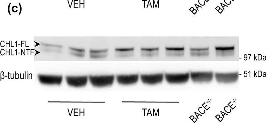
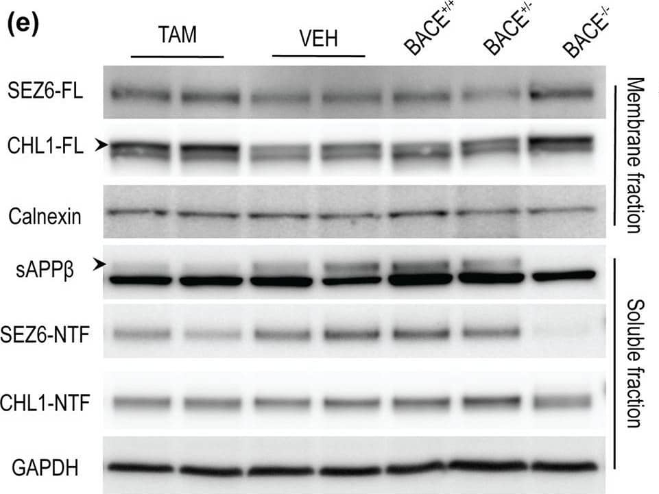
BACE1-mediated processing of APP and CHL1 is reduced in cortex of aged BACE1 cKO mice following tamoxifen treatment. Cortex homogenates from TAM- or VEH-treated mice were resolved by SDS-PAGE for Western blot analysis of APP and CHL1 processing. Homogenates from aged-matched BACE+/− and BACE1−/− were also loaded as control samples. APP-FL, pC99 and pC89 were normalized to GAPDH (MAB374) while CHL1-FL and CHL1-NTF were normalized to beta-tubulin (JDR.3B8). Protein amount was normalized to protein levels in control mice injected with vehicle (set at 1). Representative blots of (a) APP-FL (C1/6.1), (b) APP-CTFs (C1/6.1) and (c) CHL1. (d) Densitometry analysis of protein expression. APP processing was reduced in TAM-treated mice as demonstrated by the accumulation of APP-FL (C1/6.1), and reduced levels of the betaCTFs pC99 and pC89. betaCTFs were clearly identified because missing in the BACE1−/− sample. CHL1-FL (AF2147) levels were increased and CHL1-NTF levels were significantly reduced. Furthermore, the CHL1-NTF/CHL1-FL ratio was significantly decreased in TAM-treated mice demonstrating reduced BACE1 processing (VEH n = 7; TAM n = 7). (e) Quantification of A betax-40 was performed by MSD immunoassay on cortex homogenates and expressed as pMol/g of cortex. The decrease of levels of A betax-40 in TAM-treated mice was comparable to the one observed in samples collected from young TAM-treated mice (~50% decrease) (VEH n = 7; TAM n = 7). Results were plotted as Mean ± SEM, *p < 0.05; **p < 0.005; ***p < 0.001; ****p < 0.0001; n.s. = not significant, Student’s t test. Image collected and cropped by CiteAb from the following publication (https://pubmed.ncbi.nlm.nih.gov/31882662), licensed under a CC-BY license. Not internally tested by R&D Systems.Applications for Mouse CHL-1/L1CAM-2 Antibody
Immunohistochemistry
Sample: Perfusion fixed frozen sections of mouse brain (cortex)
Simple Western
Sample: Mouse brain (cortex) tissue and mouse brain stem tissue
Western Blot
Sample: Mouse brain stem tissue
Reviewed Applications
Read 4 reviews rated 4.8 using AF2147 in the following applications:
Formulation, Preparation, and Storage
Purification
Reconstitution
Formulation
Shipping
Stability & Storage
- 12 months from date of receipt, -20 to -70 °C as supplied.
- 1 month, 2 to 8 °C under sterile conditions after reconstitution.
- 6 months, -20 to -70 °C under sterile conditions after reconstitution.
Background: CHL-1/L1CAM-2
Close homolog of L1 (CHL-1), also known as cell adhesion L1-like (CALL) and L1 cell adhesion molecule 2 (L1CAM-2), belongs to the L1 subfamily of immunoglobulin (Ig) superfamily cell adhesion molecules, which also includes L1, neurofascin and NgCAM-related cell adhesion molecule (NrCAM) (1‑3). These molecules are type I transmembrane proteins that have 6 Ig-like domains and 4‑5 fibronectin type III-like (FNIII) domains in their extracellular regions. They also share a highly conserved cytoplasmic region of approximately 110 amino acid (aa) residues containing an ankyrin-binding site. CHL-1 is expressed as a highly glycosylated 185 kDa transmembrane protein by subpopulations of neurons and glia of the central and peripheral nervous system (4, 5). Ectodomain shedding via the metalloprotease-disintegrin ADAM8 releases 165 kDa and 125 kDa soluble CHL-1 fragments, which can diffuse away to function at distant sites (6). CHL-1 is not capable of homotypic interactions, but an extracellular binding partner of CHL-1 has not been identified (4). Human CHL1 has been mapped to chromosome 3p26 and is a candidate gene for 3p- syndrome characterized by mental impairment (7). A missense CHL1 polymorphism associated increased risk of schizophrenia, has also been reported (8). The functional importance of CHL-1 in the nervous system is also evident in CHL-1 deficient mice, which display behavioral abnormalities and show misguided axons within the hippocampus and olfactory tract (9). Enhanced ectodomain-shedding of CHL-1 is also observed in Wobbler mice, the neurodegenerative mutant mice (6). In vitro, soluble or substrate-coated CHL-1 promotes neurite outgrowth and neuronal survival of both cerebellar and hippocampal neurons. Cell surface CHL-1 interacts with integrins in cis to potentiate integrin-dependent cell migration toward extracellular matrix proteins (10). For this enhanced cell motility, CHL-1 linkage to the actin cytoskeleton via interaction between ankyrin and the CHL-1 cytoplasmic region is required.
References
- Moos, M. et al. (1988) Nature 334:701.
- Holm, J. et al. (1996) Eur. J. Neusci. 8:1613.
- Wei, M. et al. (1998) Hum. Genet. 103:355.
- Hillenbrand, R. et al. (1999) Eur. J. Neurosci. 11:813.
- Liu, Q. et al. (2000) J. Neurosci. 20:7682.
- Naus, S. et al. (2004) J. Biol. Chem. 279:16083.
- Angeloni, D. et al. (1999) Am. J. Med. Genet. 86:482.
- Sakurai, K. et al. (2002) Mol. Psychiatry 7:412.
- Montag-Sallaz, M. et al. (2002) Mol. Cell. Biol. 22:7967.
- Buhusi, M. et al. (2003) J. Biol. Chem. 278(27):25024.
Long Name
Alternate Names
Gene Symbol
UniProt
Additional CHL-1/L1CAM-2 Products
Product Documents for Mouse CHL-1/L1CAM-2 Antibody
Product Specific Notices for Mouse CHL-1/L1CAM-2 Antibody
For research use only


