Mouse Flt-3 Ligand/FLT3L Antibody
R&D Systems, part of Bio-Techne | Catalog # AF427


Key Product Details
Validated by
Species Reactivity
Validated:
Cited:
Applications
Validated:
Cited:
Label
Antibody Source
Product Specifications
Immunogen
Gly27-Arg188
Accession # P49772
Specificity
Clonality
Host
Isotype
Endotoxin Level
Scientific Data Images for Mouse Flt-3 Ligand/FLT3L Antibody
Cell Proliferation Induced by Flt-3 Ligand/FLT3L and Neutralization by Mouse Flt-3 Ligand/FLT3L Antibody.
Recombinant Mouse Flt-3 Ligand/FLT3L (Catalog # 427-FL) stimulates proliferation in BaF3 mouse pro-B cell line transfected with mouse Flt-3 in a dose-dependent manner (orange line). Proliferation elicited by Recombinant Mouse Flt-3 Ligand/FLT3L (10 ng/mL) is neutralized (green line) by increasing concentrations of Goat Anti-Mouse Flt-3 Ligand/FLT3L Antigen Affinity-purified Polyclonal Antibody (Catalog # AF427). The ND50 is typically 0.05-0.25 µg/mL.Flt-3 Ligand/FLT3L in HT‑2 Mouse Cell Line.
Flt-3 Ligand/FLT3L was detected in immersion fixed HT-2 mouse T cell line using Goat Anti-Mouse Flt-3 Ligand/FLT3L Antigen Affinity-purified Polyclonal Antibody (Catalog # AF427) at 5 µg/mL for 3 hours at room temperature. Cells were stained using the NorthernLights™ 557-conjugated Anti-Goat IgG Secondary Antibody (red; Catalog # NL001) and counterstained with DAPI (blue). Specific staining was localized to cytoplasm. View our protocol for Fluorescent ICC Staining of Non-adherent Cells.Detection of Mouse Flt-3 Ligand/FLT3L by Flow Cytometry
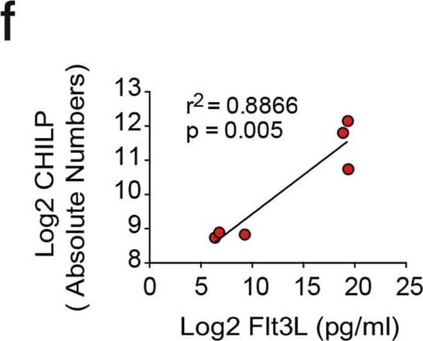
Flt3L-producing tumors do not expand mature ILCs in periphery. Mice were injected with 2 × 10⁶ B16-Flt3L or B16/PBS as a control. Two weeks after tumor injection, ILCs were analyzed by FACS in the lungs, small intestine and colonic lamina propria. (a) Representative dot plots showing NK1.1+T-bet+Eomes− ILC1, NK1.1+T-bet+Eomes+ cNK, GATA-3+ ILC2, ROR gammat+ ILC3 and NKp46+ and CCR6+ ILC3 subsets gated on CD45+ Lin (CD3, CD19)neg CD90+ cells in the colonic lamina propria and (b) quantification of absolute numbers (n = 5 to 7/group, 3 experiments). Representative dot plots (c) and absolute numbers (d) of ILC2 and ILC3 in the small intestine lamina propria and lungs of B16-Flt3L and B16/PBS injected mice (n = 5 to 8/group, 3 experiments). (e) Representative dot plots of IL22+ ROR gammat+ ILC3 and IL-5+ GATA-3+ ILC2 in the small intestine of B16-Flt3L and B16 injected mice (n = 3 to 4, 2 experiments). *P < 0.05; ns, not significant; Student’s t-test (b,d,e). Error bars represent SEM in all panels. Image collected and cropped by CiteAb from the following open publication (https://pubmed.ncbi.nlm.nih.gov/29317685), licensed under a CC-BY license. Not internally tested by R&D Systems.Applications for Mouse Flt-3 Ligand/FLT3L Antibody
Immunocytochemistry
Sample: Immersion fixed HT-2 mouse T cell line
Western Blot
Sample: Recombinant Mouse Flt-3 Ligand/FLT3L (Catalog # 427-FL)
Neutralization
Mouse Flt-3 Ligand/FLT3L Sandwich Immunoassay
Formulation, Preparation, and Storage
Purification
Reconstitution
Formulation
Shipping
Stability & Storage
- 12 months from date of receipt, -20 to -70 °C as supplied.
- 1 month, 2 to 8 °C under sterile conditions after reconstitution.
- 6 months, -20 to -70 °C under sterile conditions after reconstitution.
Background: Flt-3 Ligand/FLT3L
Flt-3 Ligand, also known as FL, is an alpha-helical cytokine that promotes the differentiation of multiple hematopoietic cell lineages (1‑3). Mature mouse Flt-3 Ligand consists of a 161 amino acid (aa) extracellular domain (ECD) with a cytokine-like domain and a juxtamembrane tether region, a 21 aa transmembrane segment, and a 22 aa cytoplasmic tail (4‑6). Within the ECD, mouse Flt-3 Ligand shares 71% and 81% aa sequence identity with human and rat Flt-3 Ligand, respectively. Mouse and human Flt-3 Ligand show cross-species activity (4, 5, 7). Flt-3 Ligand is expressed as a noncovalently-linked dimer by T cells and bone marrow and thymic fibroblasts (1, 8). Each 36 kDa chain carries approximately 12 kDa of N- and O-linked carbohydrates (8). Alternate splicing and proteolytic cleavage of the transmembrane form can generate a soluble 30 kDa fragment that includes the cytokine domain (4, 8). Alternate splicing of mouse Flt-3 Ligand also generates a membrane-associated isoform with a 57 aa substitution following the cytokine domain (4, 5, 8, 9). Both transmembrane and soluble Flt-3 Ligand signal through the tyrosine kinase receptor Flt-3/Flk-2 (3‑6). Flt-3 Ligand induces the expansion of monocytes and immature dendritic cells as well as early B cell lineage differentiation (2, 10). It synergizes with IL-3, GM-CSF, and SCF to promote the mobilization and myeloid differentiation of hematopoietic stem cells (4, 5, 7). It cooperates with IL-2, -6, -7, and -15 to induce NK cell development and with IL-3, -7, and -11 to induce terminal B cell maturation (1, 11). Animal studies also show Flt-3 Ligand to reduce the severity of experimentally induced allergic inflammation (12).
References
- Wodnar-Filipowicz, A. (2003) News Physiol. Sci. 18:247.
- Dong, J. et al. (2002) Cancer Biol. Ther. 1:486.
- Gilliland, D.G. and J.D. Griffin (2002) Blood 100:1532.
- Hannum, C. et al. (1994) Nature 368:643.
- Lyman, S.D. et al. (1993) Cell 75:1157.
- Savvides, S.N. et al. (2000) Nat. Struct. Biol. 7:486.
- Lyman, S.D. et al. (1994) Blood 83:2795.
- McClanahan, T. et al. (1996) Blood 88:3371.
- Lyman, S.D. et al. (1995) Oncogene 10:149.
- Diener, K.R. et al. (2008) Exp. Hematol. 36:51.
- Farag, S.S. and M.A. Caligiuri (2006) Blood Rev. 20:123.
- Edwan, J.H. et al. (2004) J. Immunol. 172:5016.
Long Name
Alternate Names
Gene Symbol
UniProt
Additional Flt-3 Ligand/FLT3L Products
Product Documents for Mouse Flt-3 Ligand/FLT3L Antibody
Product Specific Notices for Mouse Flt-3 Ligand/FLT3L Antibody
For research use only


