Mouse FoxC2 Antibody
R&D Systems, part of Bio-Techne | Catalog # AF6989

Key Product Details
Species Reactivity
Validated:
Cited:
Applications
Validated:
Cited:
Label
Antibody Source
Product Specifications
Immunogen
Ala412-Tyr494
Accession # Q61850
Specificity
Clonality
Host
Isotype
Scientific Data Images for Mouse FoxC2 Antibody
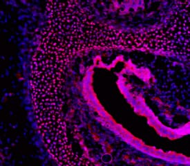
FoxC2 in Mouse Embryo.
FoxC2 was detected in immersion fixed frozen sections of mouse embryo (E15.5) using Sheep Anti-Mouse FoxC2 Antigen Affinity-purified Polyclonal Antibody (Catalog # AF6989) at 10 µg/mL overnight at 4 °C. Tissue was stained using the NorthernLights™ 557-conjugated Anti-Sheep IgG Secondary Antibody (red; Catalog # NL010) and counterstained with DAPI (blue). Specific staining was localized to periocular mesenchyme. View our protocol for Fluorescent IHC Staining of Frozen Tissue Sections.Detection of Mouse Mouse FoxC2 Antibody by Immunohistochemistry
YAP and TAZ are required for the maintenance of LVs. The lymphatic vessels in the dorsal skin of E16.5 and E18.5 control and Lyve1-Cre;Yapf/f;Tazf/f embryos were analyzed by whole-mount immunohistochemistry. (A,B) LVs were observed in the collecting lymphatic vessels of E16.5 control and Lyve1-Cre;Yapf/f;Tazf/f embryos (arrows). (C,D) The migrating front of E16.5 control (C) and Lyve1-Cre;Yapf/f;Tazf/f (D) embryos appeared comparable. (E-G) At E18.5, the lymphatic vessels from the left and right sides have merged to form a network in control embryos (E). In contrast, huge gaps were observed in between the migrating fronts of E18.5 Lyve1-Cre;Yapf/f;Tazf/f embryos (F, magenta lines). The lymphatic vessels of mutant embryos were also dilated. The distance between the migrating fronts and the diameter of vessels are quantified in G. (H,I) LVs were observed in the collecting lymphatic vessels of E18.5 control embryos (H, yellow arrows). In contrast, the dilated lymphatic vessels of E18.5 Lyve1-Cre;Yapf/f;Tazf/f embryos lacked LVs (I). The various parameters of lymphatic vascular patterning were quantified and are plotted in G. n=4 embryos per each genotype. ****P<0.0001. Data are mean±s.e.m. Scale bars: 200 µm in A-D; 500 µm in E,F; 200 µm in H,I. Image collected and cropped by CiteAb from the following publication (https://pubmed.ncbi.nlm.nih.gov/33060128), licensed under a CC-BY license. Not internally tested by R&D Systems.Detection of Mouse Mouse FoxC2 Antibody by Immunohistochemistry
YAP and TAZ are required for the maintenance of LVs. The lymphatic vessels in the dorsal skin of E16.5 and E18.5 control and Lyve1-Cre;Yapf/f;Tazf/f embryos were analyzed by whole-mount immunohistochemistry. (A,B) LVs were observed in the collecting lymphatic vessels of E16.5 control and Lyve1-Cre;Yapf/f;Tazf/f embryos (arrows). (C,D) The migrating front of E16.5 control (C) and Lyve1-Cre;Yapf/f;Tazf/f (D) embryos appeared comparable. (E-G) At E18.5, the lymphatic vessels from the left and right sides have merged to form a network in control embryos (E). In contrast, huge gaps were observed in between the migrating fronts of E18.5 Lyve1-Cre;Yapf/f;Tazf/f embryos (F, magenta lines). The lymphatic vessels of mutant embryos were also dilated. The distance between the migrating fronts and the diameter of vessels are quantified in G. (H,I) LVs were observed in the collecting lymphatic vessels of E18.5 control embryos (H, yellow arrows). In contrast, the dilated lymphatic vessels of E18.5 Lyve1-Cre;Yapf/f;Tazf/f embryos lacked LVs (I). The various parameters of lymphatic vascular patterning were quantified and are plotted in G. n=4 embryos per each genotype. ****P<0.0001. Data are mean±s.e.m. Scale bars: 200 µm in A-D; 500 µm in E,F; 200 µm in H,I. Image collected and cropped by CiteAb from the following publication (https://pubmed.ncbi.nlm.nih.gov/33060128), licensed under a CC-BY license. Not internally tested by R&D Systems.Applications for Mouse FoxC2 Antibody
Immunohistochemistry
Sample: Immersion fixed frozen sections of mouse embryo (E15.5)
Formulation, Preparation, and Storage
Purification
Reconstitution
Formulation
Shipping
Stability & Storage
- 12 months from date of receipt, -20 to -70 °C as supplied.
- 1 month, 2 to 8 °C under sterile conditions after reconstitution.
- 6 months, -20 to -70 °C under sterile conditions after reconstitution.
Background: FoxC2
FOXC2 (Forkhead box protein C2; also BF-3, FKH-14 and MFH-1) is a 64-66 kDa member of the winged helix transcription factor gene family. It is widely expressed, including in endothelium where it induces the appearance of CXCR4 and integrin beta3, two molecules essential to cell migration. High-calorie diets also induce FOXC2 in adipocytes, inducing a brown-fat like phenotype. Mouse FOXC2 is 494 amino acids (aa) in length. It contains a forkhead DNA binding domain (aa 71-148), a short poly-Arg segment (aa 162-166), one His-rich region (aa 386-395) and an Ala/Pro-rich domain (aa 396-415). There are seven potential Ser/Thr phosphorylation sites. Over aa 412-494, mouse FOXC2 shares 99% and 87% aa identity with rat and human FOXC2, respectively.
Long Name
Alternate Names
Gene Symbol
UniProt
Additional FoxC2 Products
Product Documents for Mouse FoxC2 Antibody
Product Specific Notices for Mouse FoxC2 Antibody
For research use only


