Mouse GITR/TNFRSF18 Antibody
R&D Systems, part of Bio-Techne | Catalog # MAB52412


Key Product Details
Species Reactivity
Applications
Label
Antibody Source
Product Specifications
Immunogen
Accession # O35714
Specificity
Clonality
Host
Isotype
Scientific Data Images for Mouse GITR/TNFRSF18 Antibody
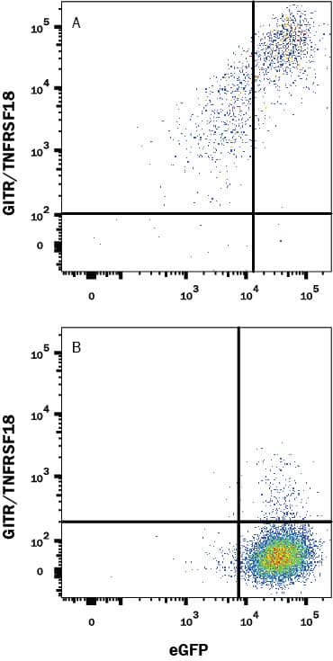
Detection of GITR/TNFRSF18 in HEK293 Human Cell Line Transfected with Mouse GITR/TNFRSF18 and eGFP by Flow Cytometry.
HEK293 human embryonic kidney cell line transfected with (A) mouse GITR/TNFRSF18 or (B) irrelevant transfectants and eGFP was stained with Rabbit Anti-Mouse GITR/TNFRSF18 Monoclonal Antibody (Catalog # MAB52412) followed by APC-conjugated Anti-Rabbit IgG Secondary Antibody (Catalog # F0111). Quadrant markers were set based on control antibody staining (Catalog # MAB1050). View our protocol for Staining Membrane-associated Proteins.GITR/TNFRSF18 in Mouse Splenocytes and NIH-3T3 Cell Line.
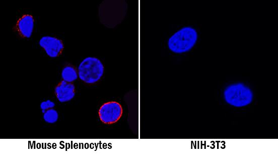
GITR/TNFRSF18 was detected in immersion fixed mouse splenocytes (left panel; positive staining) and NIH-3T3 mouse embryonic fibroblast cell line (right panel; negative staining) using Rabbit Anti-Mouse GITR/TNFRSF18 Monoclonal Antibody (Catalog # MAB52412) at 8 µg/mL for 3 hours at room temperature. Cells were stained using the NorthernLights™ 557-conjugated Anti-Rabbit IgG Secondary Antibody (red; Catalog # NL004) and counterstained with DAPI (blue). Specific staining was localized to cell surfaces. View our protocol for Fluorescent ICC Staining of Non-adherent Cells.GITR/TNFRSF18 in Mouse Spleen.
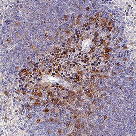
GITR/TNFRSF18 was detected in immersion fixed frozen sections of mouse spleen using Rabbit Anti-Mouse GITR/TNFRSF18 Monoclonal Antibody (Catalog # MAB52412) at 3 µg/mL for 1 hour at room temperature followed by incubation with the Anti-Rabbit IgG VisUCyte™ HRP Polymer Antibody (Catalog # VC003). Tissue was stained using DAB (brown) and counterstained with hematoxylin (blue). Specific staining was localized to lymphocytes. View our protocol for IHC Staining with VisUCyte HRP Polymer Detection Reagents.Applications for Mouse GITR/TNFRSF18 Antibody
CyTOF-ready
Flow Cytometry
Sample: HEK293 Human Cell Line Transfected with Mouse GITR/TNFRSF18 and eGFP
Immunocytochemistry
Sample: Immersion fixed mouse splenocytes and NIH-3T3 mouse embryonic fibroblast cell line
Immunohistochemistry
Sample: Immersion fixed frozen sections of mouse spleen
Formulation, Preparation, and Storage
Purification
Reconstitution
Formulation
Shipping
Stability & Storage
- 12 months from date of receipt, -20 to -70 °C as supplied.
- 1 month, 2 to 8 °C under sterile conditions after reconstitution.
- 6 months, -20 to -70 °C under sterile conditions after reconstitution.
Background: GITR/TNFRSF18
GITR (glucocorticoid-induced tumor necrosis factor receptor; also named AITR) is a member of the co‑stimulatory subset of the TNF receptor superfamily (1, 2). In mouse, the GITR gene is composed of five exons and encodes multiple length isoforms that arise from alternate splicing. The “standard”, or first reported isoform is a type I transmembrane protein, 228 amino acids (aa) in length that contains a 19 aa signal sequence, a 134 aa extracellular region, a 21 aa transmembrane segment, and a 54 aa cytoplasmic domain. The extracellular region contains four potential N-linked glycosylation sites plus three cysteine-rich pseudorepeats of about 40 aa each (3, 4). The extracellular regions of mouse and human are 57% aa identical. The cytoplasmic domain has a P-x-Q/E-E motif that is known to associate with TRAF2. This is a common characteristic of TNFRSF members with co‑stimulatory functions (4). Three other mouse GITR isoforms (B, C and D) have been reported (5). All share the same N-terminal 101 of 134 aa in the extracellular region (including pseudorepeats #1, #2 and one-half of #3). Isoform D diverges at aa #101 and continues for another 12 aa for a total length of 113 aa. This is a naturally-occurring soluble form. Isoforms B and C show splicing in their cytoplasmic tails that creates cytoplasmic domains of 118 aa and 46 aa, respectively. In both the B and C isoforms, the TRAF2 binding site is spliced out, with a p56lck binding site inserted in isoform B (4). Given its membership in the TNFRSF, it likely functions as a trimer on the cell surface (2). GITR is predominantly expressed on CD4+CD25+ regulatory T cells (Treg) and naïve CD8+ and CD4+ CD25- T cells, where its expression is up-regulated after antigen-driven activation. GITR activation provides co‑stimulatory signals for activated CD4+ CD25- T cells to enhance cell proliferation and augment cytokine production (IL-2, IL-4, IFN-gamma). On CD4+ CD25+ Treg cells, GITR activation provides co‑stimulatory signals to induce proliferation, setting Treg cells in an active/hyperproliferactive state (6‑8).
References
- Kwon, B. et al. (2003) Exp. Mol. Med. 35:8.
- Croft, M. (2003) Nat. Rev. Immunol. 3:609.
- Nocentini, G. et al. (1997) Proc. Natl. Acad. Sci. USA 94:6216.
- Nocentini, G. et al. (2000) DNA Cell Biol. 19:205.
- Nocentini, G. et al. (2000) Cell Death Differ. 7:408.
- Tone, M. et al. (2003) Proc. Natl. Acad. Sci. USA 100:15059.
- Ji, H. et al. (2004) J. Immunol. 172:5823.
- Stephens, G.L. et al. (2004) 173:5008.
Long Name
Alternate Names
Gene Symbol
UniProt
Additional GITR/TNFRSF18 Products
Product Documents for Mouse GITR/TNFRSF18 Antibody
Product Specific Notices for Mouse GITR/TNFRSF18 Antibody
For research use only

